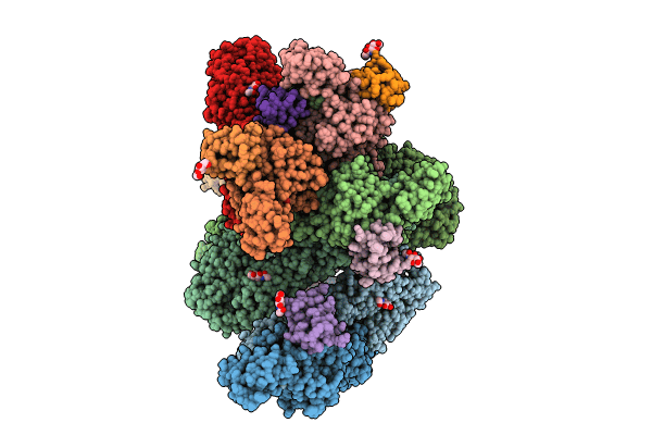
8UA4 image
Entry Detail
PDB ID:
8UA4
EMDB ID:
Title:
Structure of eastern equine encephalitis virus VLP in complex with VLDLR LA1
Biological Source:
PDB Version:
Deposition Date:
2023-09-20
Release Date:
2024-08-14
Method Details:
Experimental Method:
Resolution:
3.58 Å
Aggregation State:
PARTICLE
Reconstruction Method:
SINGLE PARTICLE
Macromolecular Entities
Polymer Type:polypeptide(L)
Description:Envelope glycoprotein E1
Chain IDs:A, D, G, J
Chain Length:441
Number of Molecules:4
Biological Source:Eastern equine encephalitis virus
Polymer Type:polypeptide(L)
Description:Envelope glycoprotein E2
Chain IDs:B, E, H, K
Chain Length:420
Number of Molecules:4
Biological Source:Eastern equine encephalitis virus
Polymer Type:polypeptide(L)
Description:Capsid protein
Mutations:K67N
Chain IDs:C, F, I, L
Chain Length:261
Number of Molecules:4
Biological Source:Eastern equine encephalitis virus
Polymer Type:polypeptide(L)
Description:Envelope glycoprotein E3
Chain IDs:M, N, O, P
Chain Length:63
Number of Molecules:4
Biological Source:Eastern equine encephalitis virus
Polymer Type:polypeptide(L)
Description:Very low-density lipoprotein receptor
Chain IDs:Q (auth: R)
Chain Length:36
Number of Molecules:1
Biological Source:Homo sapiens
Primary Citation
Structural basis for VLDLR recognition by eastern equine encephalitis virus.
Nat Commun 15 6548 6548 (2024)
PMID: 39095394 DOI: 10.1038/s41467-024-50887-9

Abstact

Eastern equine encephalitis virus (EEEV) is the most virulent alphavirus that infects humans, and many survivors develop neurological sequelae, including paralysis and intellectual disability. Alphavirus spike proteins comprise trimers of heterodimers of glycoproteins E2 and E1 that mediate binding to cellular receptors and fusion of virus and host cell membranes during entry. We recently identified very-low density lipoprotein receptor (VLDLR) and apolipoprotein E receptor 2 (ApoER2) as cellular receptors for EEEV and a distantly related alphavirus, Semliki Forest virus (SFV). Here, we use single-particle cryo-electron microscopy (cryo-EM) to determine structures of the EEEV and SFV spike glycoproteins bound to the VLDLR ligand-binding domain and found that EEEV and SFV interact with the same cellular receptor through divergent binding modes. Our studies suggest that the ability of LDLR-related proteins to interact with viral spike proteins through very small footprints with flexible binding modes results in a low evolutionary barrier to the acquisition of LDLR-related proteins as cellular receptors for diverse sets of viruses.

Legend

Protein

Chemical

Disease

Primary Citation of related structures