Deposition Date
2023-09-20
Release Date
2024-09-18
Last Version Date
2024-10-23
Entry Detail
PDB ID:
8U9X
Keywords:
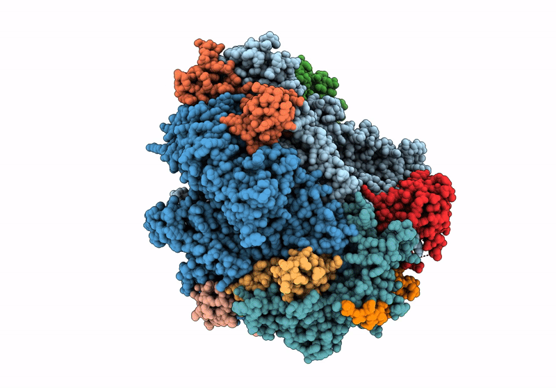
Title:
STRUCTURAL BASIS OF TRANSCRIPTION: RNA POLYMERASE II SUBSTRATE BINDING AND METAL COORDINATION AT 3.0 A OF T834P MUTANT USING A FREE-ELECTRON LASER
Biological Source:
Source Organism(s):
synthetic construct (Taxon ID: 32630)
Saccharomyces cerevisiae (Taxon ID: 4932)
Saccharomyces cerevisiae (Taxon ID: 4932)
Method Details:
Experimental Method:
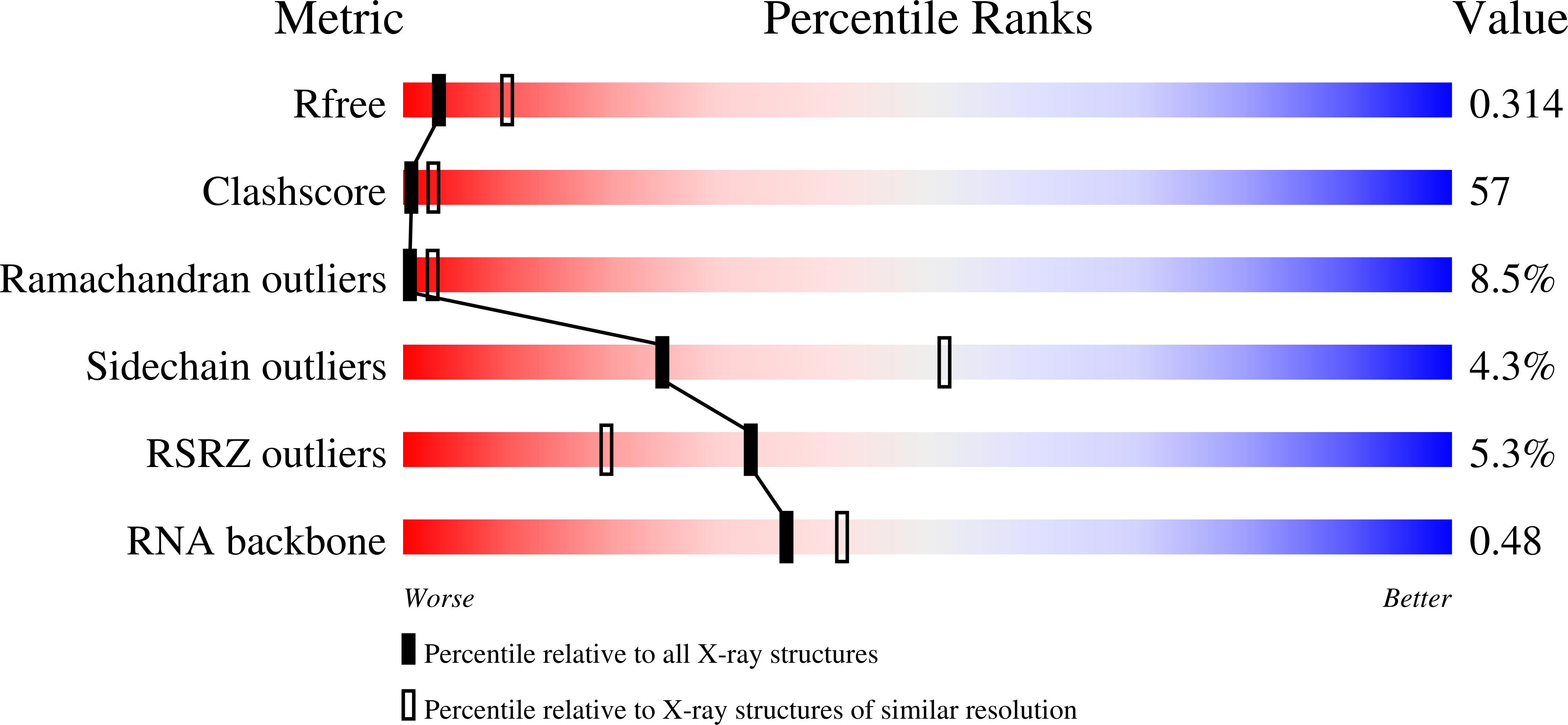
Resolution:
3.05 Å
R-Value Free:
0.30
R-Value Work:
0.28
R-Value Observed:
0.28
Space Group:
C 2 2 21