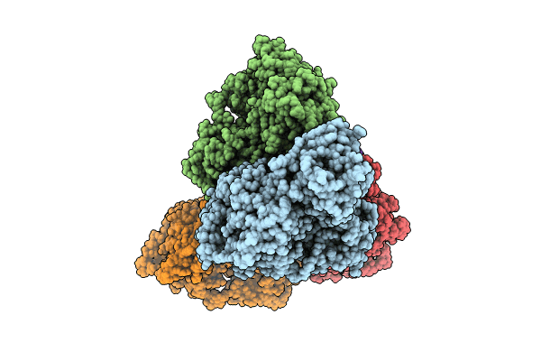
8U61 image
Deposition Date 2023-09-13
Release Date 2024-01-31
Last Version Date 2024-03-27
Entry Detail
PDB ID:
8U61
Title:
Human RADX tetramer bound to ssDNA
Biological Source:
Source Organism:
Homo sapiens (Taxon ID: 9606)
Host Organism:
Method Details:
Experimental Method:
Resolution:
4.00 Å
Aggregation State:
PARTICLE
Reconstruction Method:
SINGLE PARTICLE
Macromolecular Entities
Structures with similar UniProt ID
Protein Blast
Polymer Type:polypeptide(L)
Molecule:RPA-related protein RADX
Gene (Uniprot):RADX
Chain IDs:A (auth: D), B, C (auth: A), D (auth: C)
Chain Length:855
Number of Molecules:4
Biological Source:Homo sapiens
Polymer Type:polydeoxyribonucleotide
Molecule:dT25 DNA (25-MER)
Chain IDs:E
Chain Length:25
Number of Molecules:1
Biological Source:Homo sapiens
Ligand Molecules
Primary Citation
Structure of RADX and mechanism for regulation of RAD51 nucleofilaments.
Proc.Natl.Acad.Sci.USA 121 e2316491121 e2316491121 (2024)
PMID: 38466836 DOI: 10.1073/pnas.2316491121

Abstact

Replication fork reversal is a fundamental process required for resolution of encounters with DNA damage. A key step in the stabilization and eventual resolution of reversed forks is formation of RAD51 nucleoprotein filaments on exposed single strand DNA (ssDNA). To avoid genome instability, RAD51 filaments are tightly controlled by a variety of positive and negative regulators. RADX (RPA-related RAD51-antagonist on the X chromosome) is a recently discovered negative regulator that binds tightly to ssDNA, directly interacts with RAD51, and regulates replication fork reversal and stabilization in a context-dependent manner. Here, we present a structure-based investigation of RADX's mechanism of action. Mass photometry experiments showed that RADX forms multiple oligomeric states in a concentration-dependent manner, with a predominance of trimers in the presence of ssDNA. The structure of RADX, which has no structurally characterized orthologs, was determined ab initio by cryo-electron microscopy (cryo-EM) from maps in the 2 to 4 Å range. The structure reveals the molecular basis for RADX oligomerization and the coupled multi-valent binding of ssDNA binding. The interaction of RADX with RAD51 filaments was imaged by negative stain EM, which showed a RADX oligomer at the end of filaments. Based on these results, we propose a model in which RADX functions by capping and restricting the end of RAD51 filaments.

Legend

Protein

Chemical

Disease

Primary Citation of related structures