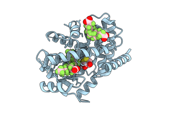
8U57 image
Entry Detail
PDB ID:
8U57
Keywords:
Title:
PPARg LBD in complex with perfluorooctanoic acid (PFOA)
Biological Source:
Source Organism:
PDB Version:
Deposition Date:
2023-09-12
Release Date:
2024-07-24
Method Details:
Experimental Method:
Resolution:
2.00 Å
R-Value Free:
0.25
R-Value Work:
0.22
R-Value Observed:
0.22
Space Group:
P 43 21 2
Macromolecular Entities
Polymer Type:polypeptide(L)
Description:Peroxisome proliferator-activated receptor gamma
Chain IDs:A
Chain Length:290
Number of Molecules:1
Biological Source:Homo sapiens
Modified Residue
Compound ID Chain ID Parent Comp ID Details 2D Image
CSO A CYS modified residue
Primary Citation
A structural basis for the activation of peroxisome proliferator-activated receptor gamma (PPAR gamma ) by perfluorooctanoic acid (PFOA).
Chemosphere 354 141723 141723 (2024)
PMID: 38494006 DOI: 10.1016/j.chemosphere.2024.141723

Abstact

Perfluorooctanoic acid (PFOA) is a widespread environmental pollutant of the perfluoroalkyl substance (PFAS) class that is extremely resistant to environmental and metabolic degradation, leading to bioaccumulation. PFOA exposure has been linked to many health effects including endocrine disruption and metabolic dysregulation, but our understanding of the molecular mechanisms resulting in these outcomes remains incomplete. One target affected by PFOA is the ligand regulated nuclear receptor peroxisome proliferator-activated receptor gamma (PPARγ) which plays a critical role in controlling metabolic homeostasis through regulating processes such as adipogenesis, glucose homeostasis, inflammation and osteogenesis. It has been previously established that PFOA activates PPARγ through binding to the PPARγ ligand binding domain (PPARγ LBD) leading to increased expression of PPARγ controlled target genes. However, the mechanism by which PFOA achieves this has remained elusive. Here, we employed a combination of X-ray crystallography and fluorescence polarization assays to provide a structural basis for PFOA mediated activation of PPARγ via binding to the PPARγ LBD. Using X-ray crystallography, the cocrystal structure of the PPARγ LBD:PFOA complex was solved. This revealed that PFOA occupies three distinct sites, two within the PPARγ LBD and one within the activation function 2 (AF2) on the protein surface. Structural comparison of PFOA binding with previously reported PPARγ:ligand complexes supports that PFOA activates PPARγ by a partial agonist mechanism at micromolar concentrations. Fluorescence polarization assays also revealed that PFOA binding to the AF2 is unlikely to occur in a cellular context and confirmed that PFOA behaves as a partial agonist in vitro, weakly recruiting a coactivator peptide to the AF2 of the PPARγ LBD. This discovery provides an advancement in understanding PFOA mediated regulation of PPARγ, giving new insight regarding regulation of PPARγ by PFAS and PFAS substitutes in general and can be applied to the design and assessment of safer PFAS.

Legend

Protein

Chemical

Disease

Primary Citation of related structures