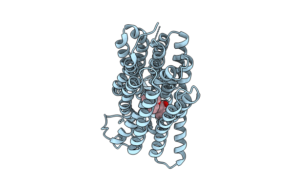
8U3H image
Entry Detail
PDB ID:
8U3H
EMDB ID:
Title:
Structure of Fmoc-Leu-OH bound Sialin
Biological Source:
Source Organism:
Host Organism:
PDB Version:
Deposition Date:
2023-09-07
Release Date:
2024-05-29
Method Details:
Experimental Method:
Resolution:
3.67 Å
Aggregation State:
PARTICLE
Reconstruction Method:
SINGLE PARTICLE
Macromolecular Entities
Polymer Type:polypeptide(L)
Description:Sialin
Chain IDs:A
Chain Length:503
Number of Molecules:1
Biological Source:Homo sapiens
Primary Citation
Structure and inhibition of the human lysosomal transporter Sialin.
Nat Commun 15 4386 4386 (2024)
PMID: 38782953 DOI: 10.1038/s41467-024-48535-3

Abstact

Sialin, a member of the solute carrier 17 (SLC17) transporter family, is unique in its ability to transport not only sialic acid using a pH-driven mechanism, but also transport mono and diacidic neurotransmitters, such as glutamate and N-acetylaspartylglutamate (NAAG), into synaptic vesicles via a membrane potential-driven mechanism. While most transporters utilize one of these mechanisms, the structural basis of how Sialin transports substrates using both remains unclear. Here, we present the cryogenic electron-microscopy structures of human Sialin: apo cytosol-open, apo lumen-open, NAAG-bound, and inhibitor-bound. Our structures show that a positively charged cytosol-open vestibule accommodates either NAAG or the Sialin inhibitor Fmoc-Leu-OH, while its luminal cavity potentially binds sialic acid. Moreover, functional analyses along with molecular dynamics simulations identify key residues in binding sialic acid and NAAG. Thus, our findings uncover the essential conformational states in NAAG and sialic acid transport, demonstrating a working model of SLC17 transporters.

Legend

Protein

Chemical

Disease

Primary Citation of related structures