Deposition Date
2023-09-07
Release Date
2025-04-09
Last Version Date
2025-10-15
Entry Detail
PDB ID:
8U38
Keywords:
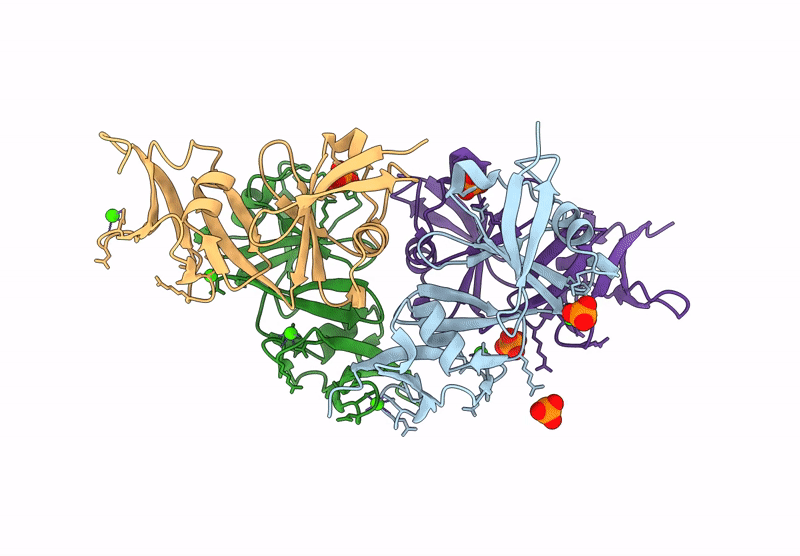
Title:
Structure of a bacterial multi-ubiquitin domain protein
Biological Source:
Source Organism(s):
Methylobacterium brachiatum (Taxon ID: 269660)
Expression System(s):
Method Details:
Experimental Method:
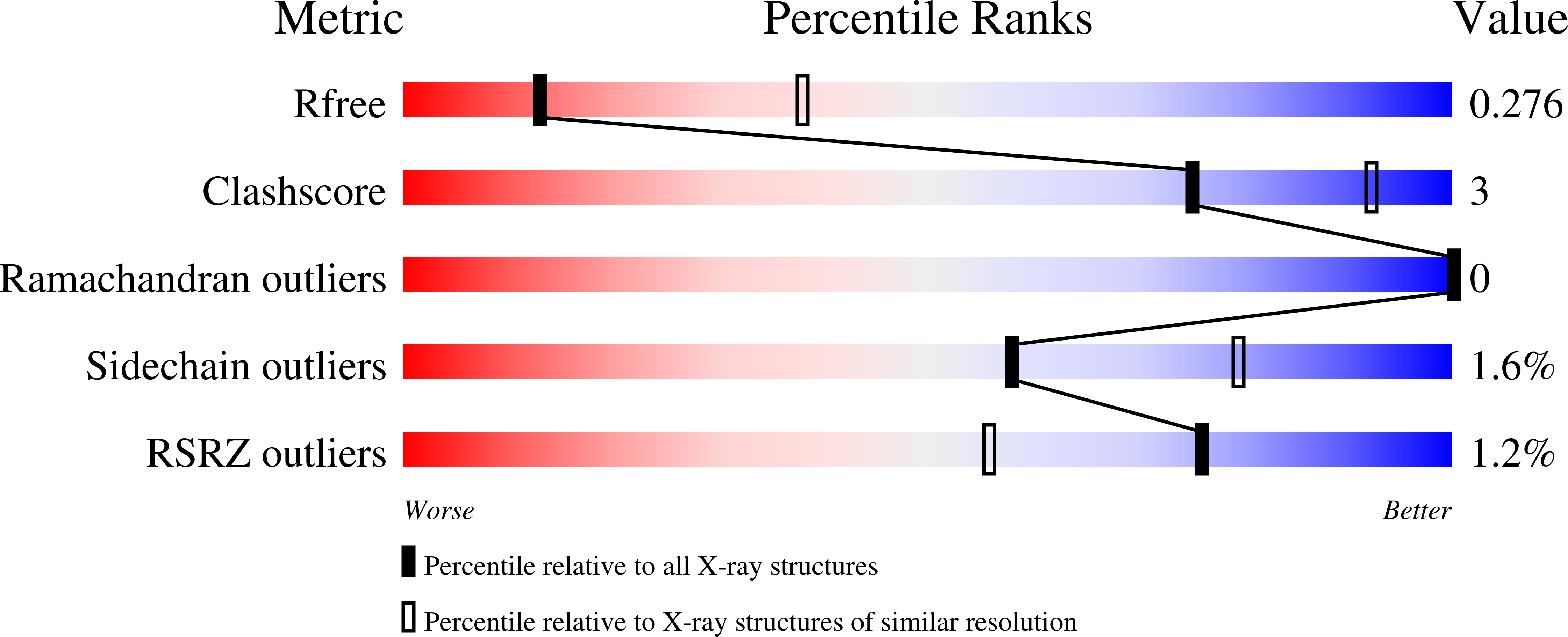
Resolution:
3.04 Å
R-Value Free:
0.27
R-Value Work:
0.23
R-Value Observed:
0.23
Space Group:
P 31 2 1