Deposition Date
2023-08-29
Release Date
2024-09-04
Last Version Date
2025-04-09
Entry Detail
PDB ID:
8U0R
Keywords:
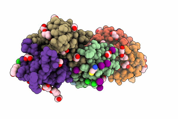
Title:
The crystal structure of protein A21, a component of the conserved poxvirus entry-fusion complex
Biological Source:
Source Organism(s):
Vaccinia virus Western Reserve (Taxon ID: 696871)
Expression System(s):
Method Details:
Experimental Method:
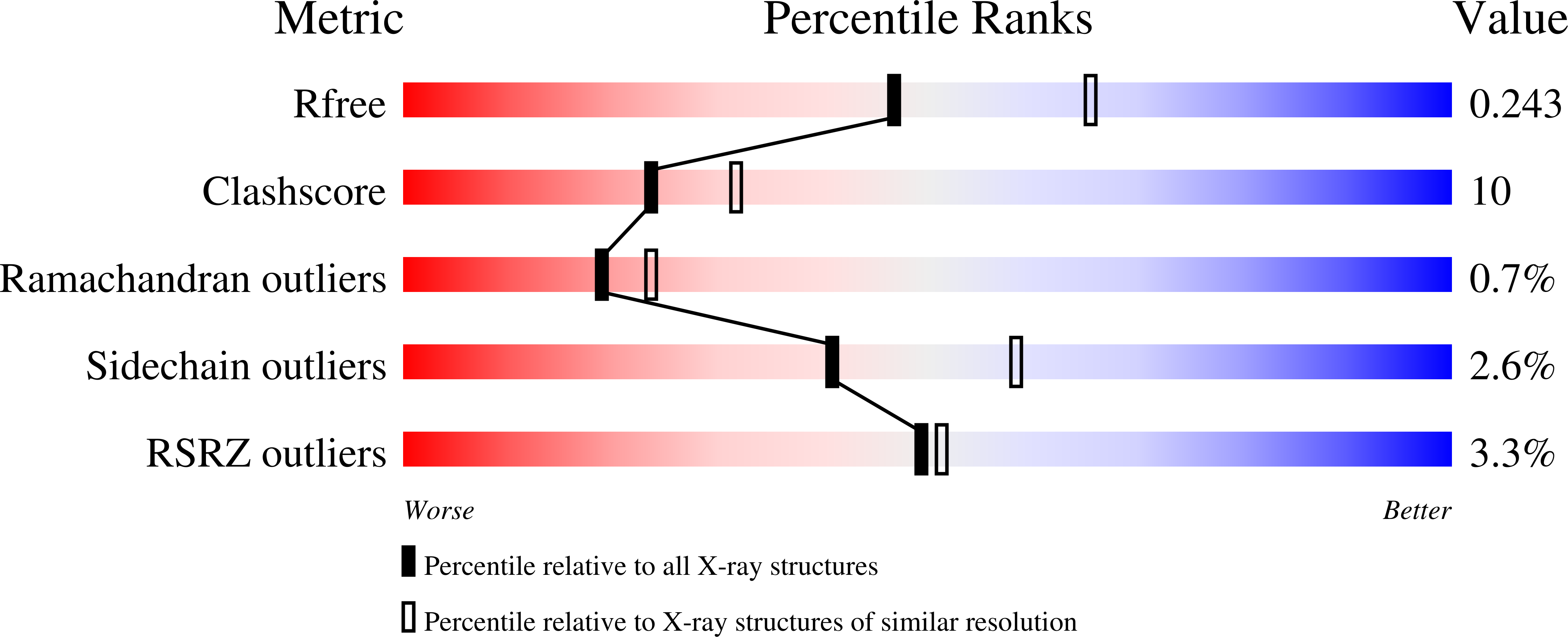
Resolution:
2.30 Å
R-Value Free:
0.23
R-Value Work:
0.21
R-Value Observed:
0.21
Space Group:
P 1 21 1