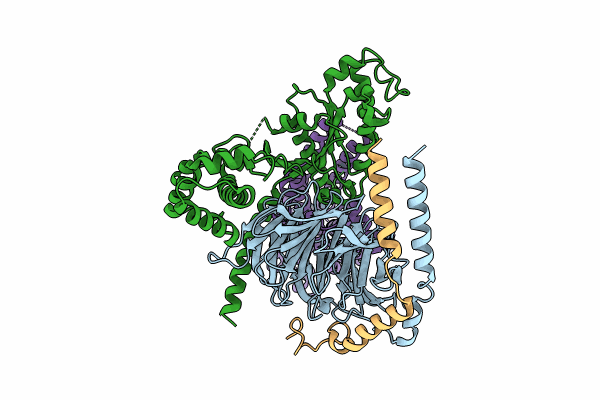
8U02 image
Entry Detail
PDB ID:
8U02
EMDB ID:
Title:
CryoEM structure of D2 dopamine receptor in complex with GoA KE mutant and dopamine
Biological Source:
Source Organism:
Host Organism:
PDB Version:
Deposition Date:
2023-08-28
Release Date:
2024-08-21
Method Details:
Experimental Method:
Resolution:
3.28 Å
Aggregation State:
PARTICLE
Reconstruction Method:
SINGLE PARTICLE
Macromolecular Entities
Polymer Type:polypeptide(L)
Description:Guanine nucleotide-binding protein G(I)/G(S)/G(T) subunit beta-1
Chain IDs:C (auth: A)
Chain Length:358
Number of Molecules:1
Biological Source:Homo sapiens
Polymer Type:polypeptide(L)
Description:Guanine nucleotide-binding protein G(o) subunit alpha
Chain IDs:B
Chain Length:354
Number of Molecules:1
Biological Source:Homo sapiens
Polymer Type:polypeptide(L)
Description:Guanine nucleotide-binding protein G(I)/G(S)/G(O) subunit gamma-2
Chain IDs:D (auth: C)
Chain Length:71
Number of Molecules:1
Biological Source:Homo sapiens
Polymer Type:polypeptide(L)
Description:D(2) dopamine receptor
Chain IDs:A (auth: R)
Chain Length:443
Number of Molecules:1
Biological Source:Homo sapiens
Ligand Molecules
Primary Citation
A neurodevelopmental disorder mutation locks G proteins in the transitory pre-activated state.
Nat Commun 15 6643 6643 (2024)
PMID: 39103320 DOI: 10.1038/s41467-024-50964-z

Abstact

Many neurotransmitter receptors activate G proteins through exchange of GDP for GTP. The intermediate nucleotide-free state has eluded characterization, due largely to its inherent instability. Here we characterize a G protein variant associated with a rare neurological disorder in humans. GαoK46E has a charge reversal that clashes with the phosphate groups of GDP and GTP. As anticipated, the purified protein binds poorly to guanine nucleotides yet retains wild-type affinity for G protein βγ subunits. In cells with physiological concentrations of nucleotide, GαoK46E forms a stable complex with receptors and Gβγ, impeding effector activation. Further, we demonstrate that the mutant can be easily purified in complex with dopamine-bound D2 receptors, and use cryo-electron microscopy to determine the structure, including both domains of Gαo, without nucleotide or stabilizing nanobodies. These findings reveal the molecular basis for the first committed step of G protein activation, establish a mechanistic basis for a neurological disorder, provide a simplified strategy to determine receptor-G protein structures, and a method to detect high affinity agonist binding in cells.

Legend

Protein

Chemical

Disease

Primary Citation of related structures