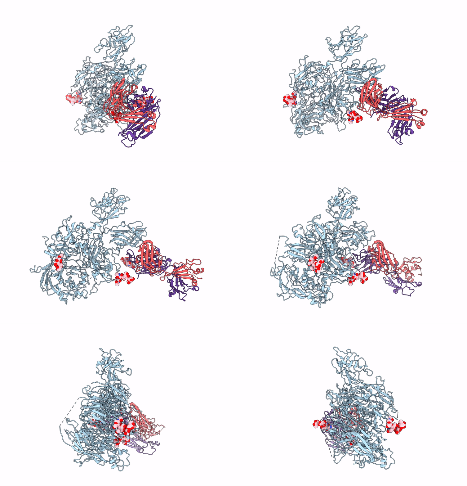
8TY1 image
Entry Detail
PDB ID:
8TY1
EMDB ID:
Title:
Cryo-EM structure of coagulation factor VIII bound to NB2E9
Biological Source:
Source Organism:
Host Organism:
PDB Version:
Deposition Date:
2023-08-24
Release Date:
2023-09-13
Method Details:
Experimental Method:
Resolution:
3.46 Å
Aggregation State:
PARTICLE
Reconstruction Method:
SINGLE PARTICLE
Macromolecular Entities
Polymer Type:polypeptide(L)
Description:Coagulation factor VIII
Chain IDs:A
Chain Length:1467
Number of Molecules:1
Biological Source:Sus scrofa, Homo sapiens
Polymer Type:polypeptide(L)
Description:NB2E9 light chain
Chain IDs:B
Chain Length:216
Number of Molecules:1
Biological Source:Homo sapiens
Polymer Type:polypeptide(L)
Description:NB2E9 heavy chain
Chain IDs:C
Chain Length:235
Number of Molecules:1
Biological Source:Homo sapiens
Ligand Molecules
Primary Citation
Structural basis for inhibition of coagulation factor VIII reveals a shared antigenic hotspot on the C1 domain.
J.Thromb.Haemost. 22 2449 2459 (2024)
PMID: 38849084 DOI: 10.1016/j.jtha.2024.05.024

Abstact

BACKGROUND Hemophilia A arises from dysfunctional or deficient coagulation factor (F)VIII and leads to inefficient fibrin clot formation and uncontrolled bleeding events. The development of antibody inhibitors is a clinical complication in hemophilia A patients receiving FVIII replacement therapy. LE2E9 is an anti-C1 domain inhibitor previously isolated from a mild/moderate hemophilia A patient and disrupts FVIII interactions with von Willebrand factor and FIXa, though the intermolecular contacts that underpin LE2E9-mediated FVIII neutralization are undefined. OBJECTIVES To determine the structure of the complex between FVIII and LE2E9 and characterize its mechanism of inhibition. METHODS FVIII was bound to the antigen binding fragment (Fab) of NB2E9, a recombinant construct of LE2E9, and its structure was determined by cryogenic electron microscopy. RESULTS This report communicates the 3.46 Å structure of FVIII bound to NB2E9, with its epitope comprising FVIII residues S2040 to Y2043, K2065 to W2070, and R2150 to H2155. Structural analysis reveals that the LE2E9 epitope overlaps with portions of the epitope for 2A9, a murine-derived inhibitor, suggesting that these residues represent a shared antigenic region on the C1 domain between FVIII-/- mice and hemophilia A patients. Furthermore, the FVIII:NB2E9 structure elucidates the orientation of the LE2E9 glycan, illustrating how the glycan sterically blocks interactions between the FVIII C1 domain and the von Willebrand factor D' domain. A putative model of the FVIIIa:FIXa complex suggests potential clashing between the NB2E9 glycan and FIXa light chain. CONCLUSION These results describe an antigenic "hotspot" on the FVIII C1 domain and provide a structural basis for engineering FVIII replacement therapeutics with reduced antigenicity.

Legend

Protein

Chemical

Disease

Primary Citation of related structures