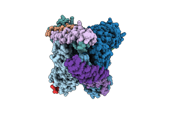
8TXR image
Deposition Date 2023-08-24
Release Date 2024-01-31
Last Version Date 2025-05-14
Entry Detail
PDB ID:
8TXR
Title:
E. coli ExoVII(H238A)
Biological Source:
Source Organism:
Escherichia coli (Taxon ID: 562)
Method Details:
Experimental Method:
Resolution:
3.80 Å
Aggregation State:
PARTICLE
Reconstruction Method:
SINGLE PARTICLE
Macromolecular Entities
Polymer Type:polypeptide(L)
Molecule:Exodeoxyribonuclease 7 large subunit
Gene (Uniprot):xseA
Mutations:H238A
Chain IDs:A, B (auth: C), I (auth: B), L (auth: D)
Chain Length:456
Number of Molecules:4
Biological Source:Escherichia coli
Polymer Type:polypeptide(L)
Molecule:Exodeoxyribonuclease 7 small subunit
Gene (Uniprot):xseB
Chain IDs:C (auth: i), D (auth: c), E (auth: g), F (auth: h), G (auth: a), H (auth: b), J (auth: o), K (auth: p), M (auth: m), N (auth: n), O (auth: j), P (auth: k), Q (auth: l), R (auth: d), S (auth: e), T (auth: f)
Chain Length:456
Number of Molecules:16
Biological Source:Escherichia coli
Ligand Molecules
Primary Citation
Structure of Escherichia coli exonuclease VII.
Proc.Natl.Acad.Sci.USA 121 e2319644121 e2319644121 (2024)
PMID: 38271335 DOI: 10.1073/pnas.2319644121

Abstact

Exonuclease VII (ExoVII) is a ubiquitous bacterial nuclease. Encoded by the xseA and xseB genes, ExoVII participates in multiple nucleic acid-dependent pathways including the processing of multicopy single-stranded DNA and the repair of covalent DNA-protein crosslinks (DPCs). Although many biochemical properties of ExoVII have been defined, little is known about its structure/function relationships. Here, we use cryoelectron microscopy (cryoEM) to determine that Escherichia coli ExoVII comprises a highly elongated XseA4·XseB24 holo-complex. Each XseA subunit dimerizes through a central extended α-helical segment decorated by six XseB subunits and a C-terminal, domain-swapped β-barrel element; two XseA2·XseB12 subcomplexes further associate using N-terminal OB (oligonucleotide/oligosaccharide-binding) folds and catalytic domains to form a spindle-shaped, catenated octaicosamer. The catalytic domains of XseA, which adopt a nuclease fold related to 3-dehydroquinate dehydratases, are sequestered in the center of the complex and accessible only through large pores formed between XseA tetramers. The architectural organization of ExoVII, combined with biochemical studies, indicate that substrate selectivity is controlled by steric access to its nuclease elements and that tetramer dissociation results from substrate DNA binding. Despite a lack of sequence and fold homology, the physical organization of ExoVII is reminiscent of Mre11·Rad50/SbcCD ATP (adenosine triphosphate)-dependent nucleases used in the repair of double-stranded DNA breaks, including those formed by DPCs through aberrant topoisomerase activity, suggesting that there may have been convergent evolutionary pressure to contend with such damage events.

Legend

Protein

Chemical

Disease

Primary Citation of related structures