Deposition Date
2023-08-04
Release Date
2023-11-29
Last Version Date
2024-03-13
Entry Detail
PDB ID:
8TP8
Keywords:
Title:
Structure of the C. crescentus WYL-activator, DriD, bound to ssDNA and cognate DNA
Biological Source:
Source Organism(s):
Caulobacter vibrioides NA1000 (Taxon ID: 565050)
Caulobacter vibrioides (Taxon ID: 155892)
Caulobacter vibrioides (Taxon ID: 155892)
Expression System(s):
Method Details:
Experimental Method:
Resolution:
2.74 Å
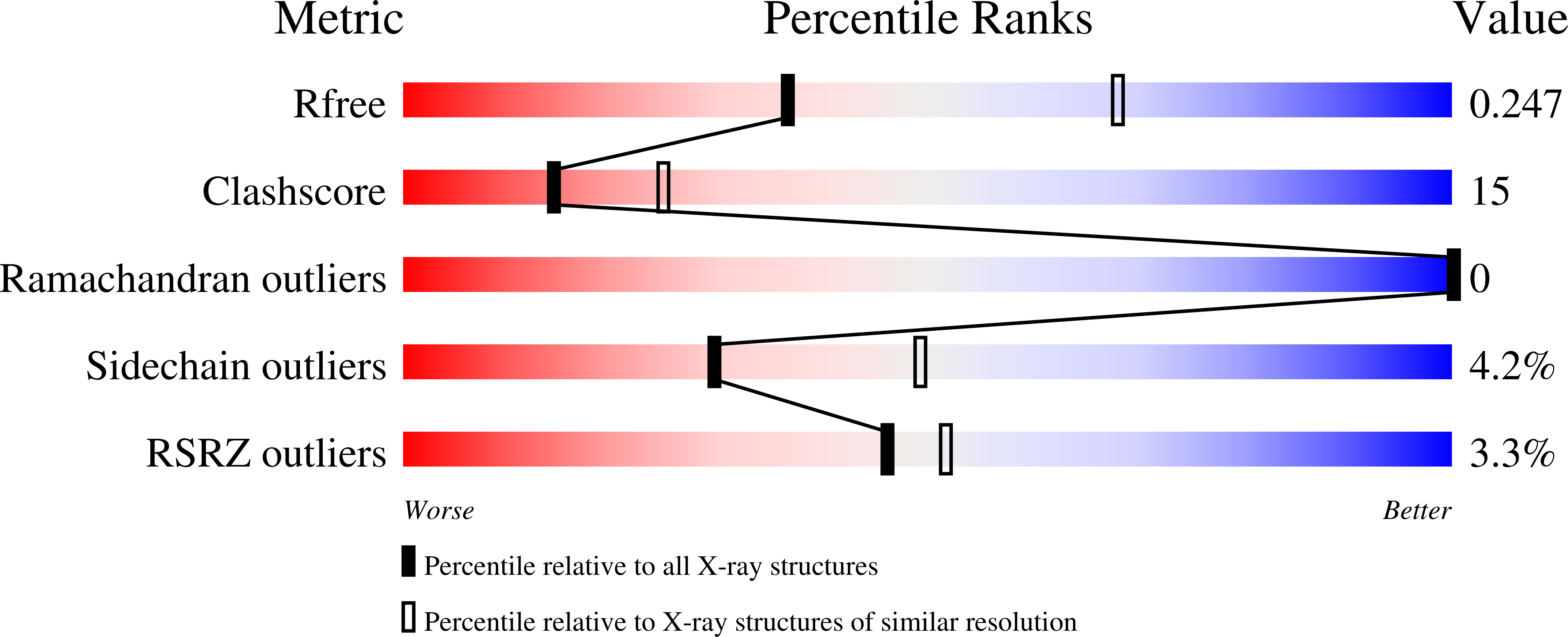
R-Value Free:
0.24
R-Value Work:
0.19
R-Value Observed:
0.19
Space Group:
P 1 21 1