Deposition Date
2023-08-02
Release Date
2023-11-15
Last Version Date
2024-05-15
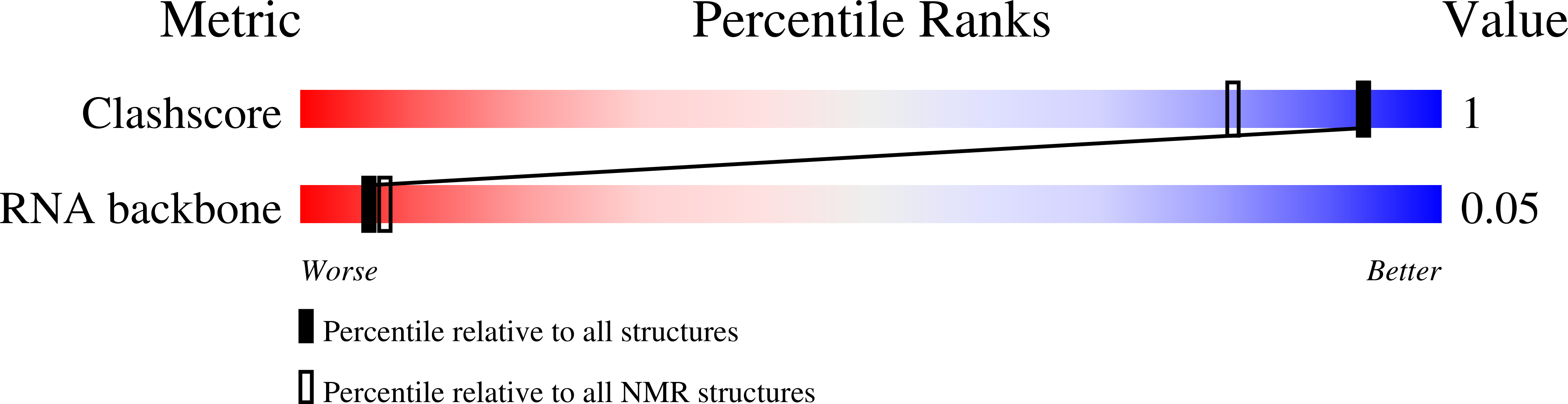
Method Details:
Experimental Method:
Conformers Calculated:
100
Conformers Submitted:
20
Selection Criteria:
structures with the least restraint violations