8TJ3 image
Entry Detail
PDB ID:
8TJ3
EMDB ID:
Title:
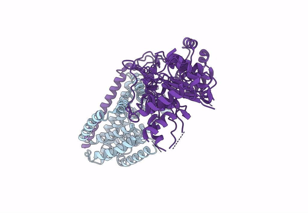
Structural basis of peptidoglycan synthesis by E. coli RodA-PBP2 complex
Biological Source:
Source Organism:
Host Organism:
PDB Version:
Deposition Date:
2023-07-20
Release Date:
2023-08-30
Method Details:
Experimental Method:
Resolution:
3.20 Å
Aggregation State:
PARTICLE
Reconstruction Method:
SINGLE PARTICLE
Macromolecular Entities
Polymer Type:polypeptide(L)
Description:Peptidoglycan glycosyltransferase MrdB
Chain IDs:A (auth: B)
Chain Length:370
Number of Molecules:1
Biological Source:Escherichia coli
Polymer Type:polypeptide(L)
Description:Peptidoglycan D,D-transpeptidase MrdA
Chain IDs:B (auth: C)
Chain Length:633
Number of Molecules:1
Biological Source:Escherichia coli
Ligand Molecules
Primary Citation

Abstact

Peptidoglycan (PG) is an essential structural component of the bacterial cell wall that is synthetized during cell division and elongation. PG forms an extracellular polymer crucial for cellular viability, the synthesis of which is the target of many antibiotics. PG assembly requires a glycosyltransferase (GT) to generate a glycan polymer using a Lipid II substrate, which is then crosslinked to the existing PG via a transpeptidase (TP) reaction. A Shape, Elongation, Division and Sporulation (SEDS) GT enzyme and a Class B Penicillin Binding Protein (PBP) form the core of the multi-protein complex required for PG assembly. Here we used single particle cryo-electron microscopy to determine the structure of a cell elongation-specific E. coli RodA-PBP2 complex. We combine this information with biochemical, genetic, spectroscopic, and computational analyses to identify the Lipid II binding sites and propose a mechanism for Lipid II polymerization. Our data suggest a hypothesis for the movement of the glycan strand from the Lipid II polymerization site of RodA towards the TP site of PBP2, functionally linking these two central enzymatic activities required for cell wall peptidoglycan biosynthesis.

Legend

Protein

Chemical

Disease

Primary Citation of related structures