Deposition Date
2023-07-04
Release Date
2023-11-22
Last Version Date
2023-12-06
Entry Detail
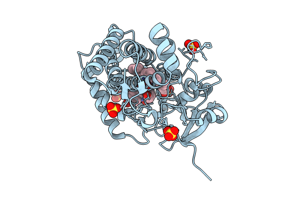
PDB ID:
8TDP
Keywords:
Title:
Time-resolved SFX-XFEL crystal structure of CYP121 bound with cYY reacted with peracetic acid for 200 milliseconds
Biological Source:
Source Organism(s):
Mycobacterium tuberculosis H37Rv (Taxon ID: 83332)
Expression System(s):
Method Details:
Experimental Method:
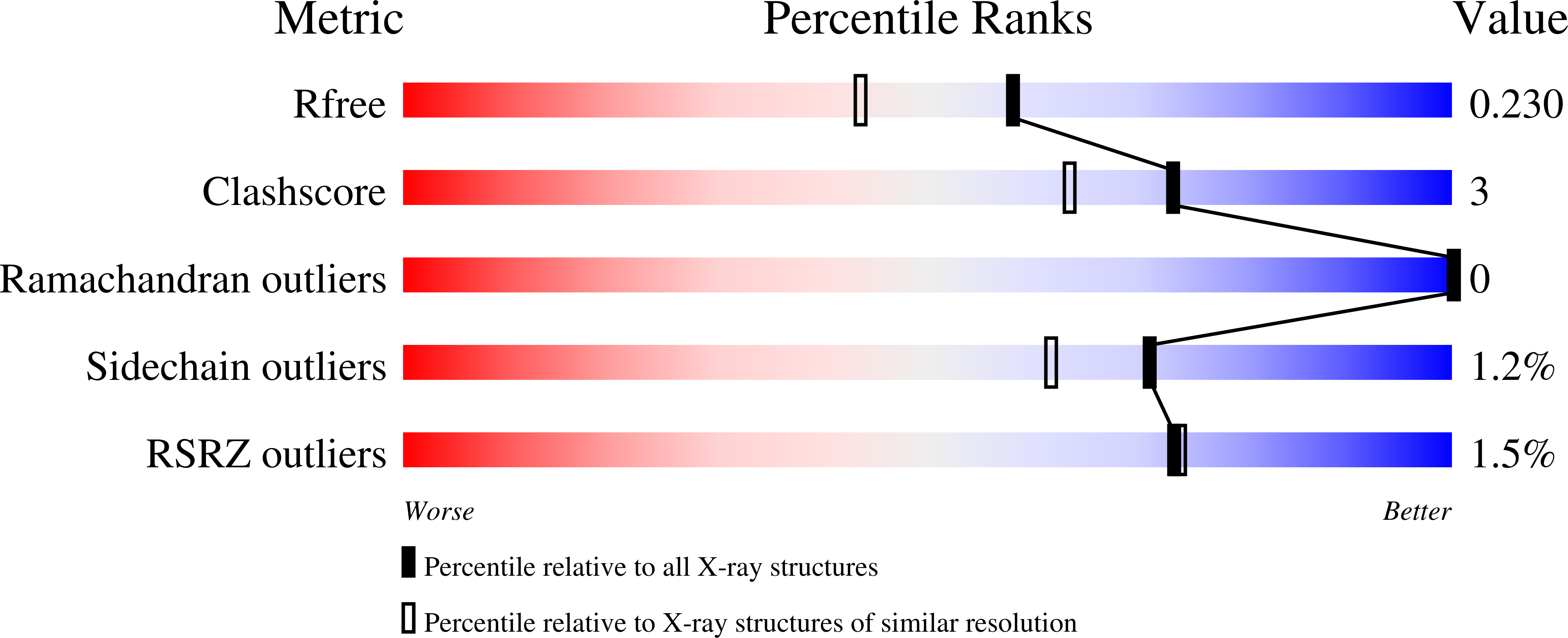
Resolution:
1.85 Å
R-Value Free:
0.22
R-Value Work:
0.18
R-Value Observed:
0.18
Space Group:
P 65 2 2