8TB2 image
Deposition Date 2023-06-28
Release Date 2024-05-08
Last Version Date 2024-05-08
Entry Detail
PDB ID:
8TB2
Keywords:
Title:
Structure of SasG (type II) (residues 165-421) from Staphylococcus aureus MW2
Biological Source:
Source Organism:
Method Details:
Experimental Method:
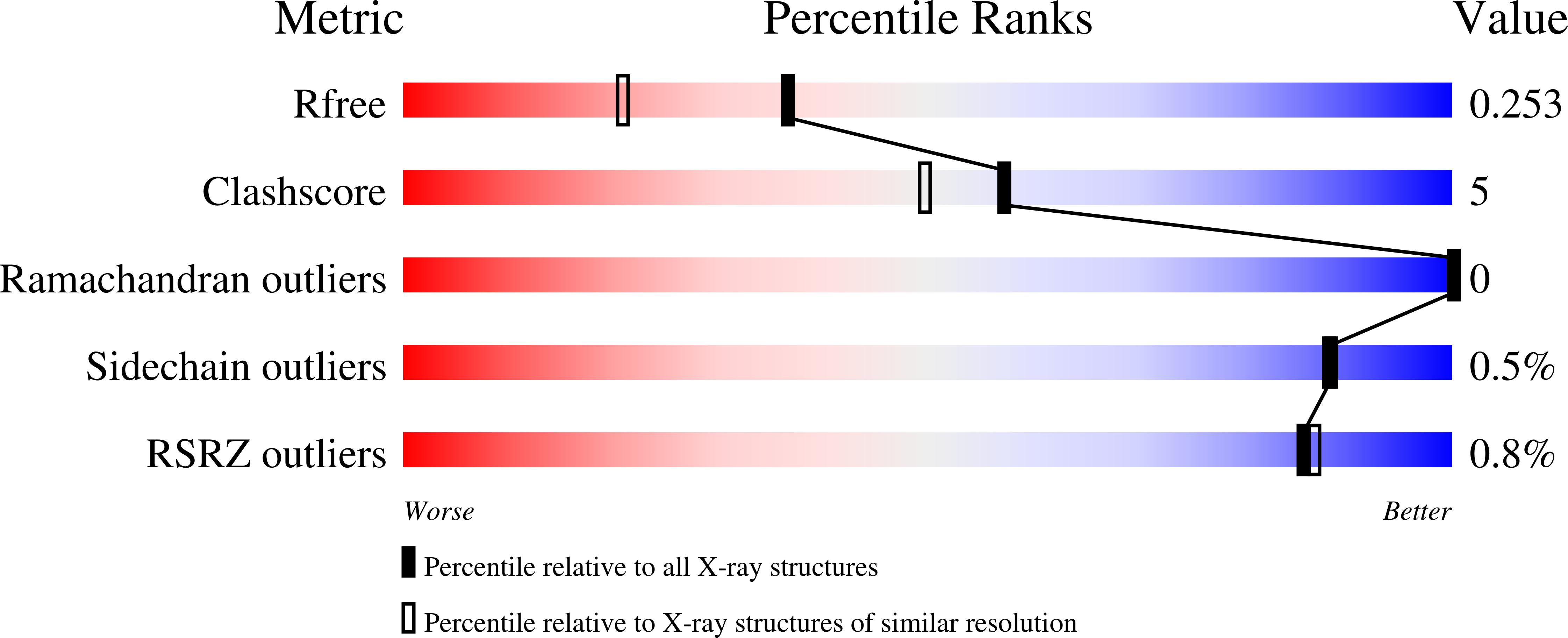
Resolution:
1.88 Å
R-Value Free:
0.25
R-Value Work:
0.18
Space Group:
P 21 21 2
Macromolecular Entities
Polymer Type:polypeptide(L)
Molecule:Putative surface protein MW2416
Gene (Uniprot):MW2416
Chain IDs:A
Chain Length:262
Number of Molecules:1
Biological Source:Staphylococcus aureus subsp. aureus MW2
Primary Citation
Staphylococcus aureus skin colonization is mediated by SasG lectin variation.
Cell Rep 43 114022 114022 (2024)
PMID: 38568806 DOI: 10.1016/j.celrep.2024.114022

Abstact

Staphylococcus aureus causes the majority of skin and soft tissue infections, but this pathogen only transiently colonizes healthy skin. However, this transient skin exposure enables S. aureus to transition to infection. The initial adhesion of S. aureus to skin corneocytes is mediated by surface protein G (SasG). Here, phylogenetic analyses reveal the presence of two major divergent SasG alleles in S. aureus: SasG-I and SasG-II. Structural analyses of SasG-II identify a nonaromatic arginine in the binding pocket of the lectin subdomain that mediates adhesion to corneocytes. Atomic force microscopy and corneocyte adhesion assays indicate that SasG-II can bind to a broader variety of ligands than SasG-I. Glycosidase treatment results in different binding profiles between SasG-I and SasG-II on skin cells. In addition, SasG-mediated adhesion is recapitulated using differentiated N/TERT keratinocytes. Our findings indicate that SasG-II has evolved to adhere to multiple ligands, conferring a distinct advantage to S. aureus during skin colonization.

Legend

Protein

Chemical

Disease

Primary Citation of related structures