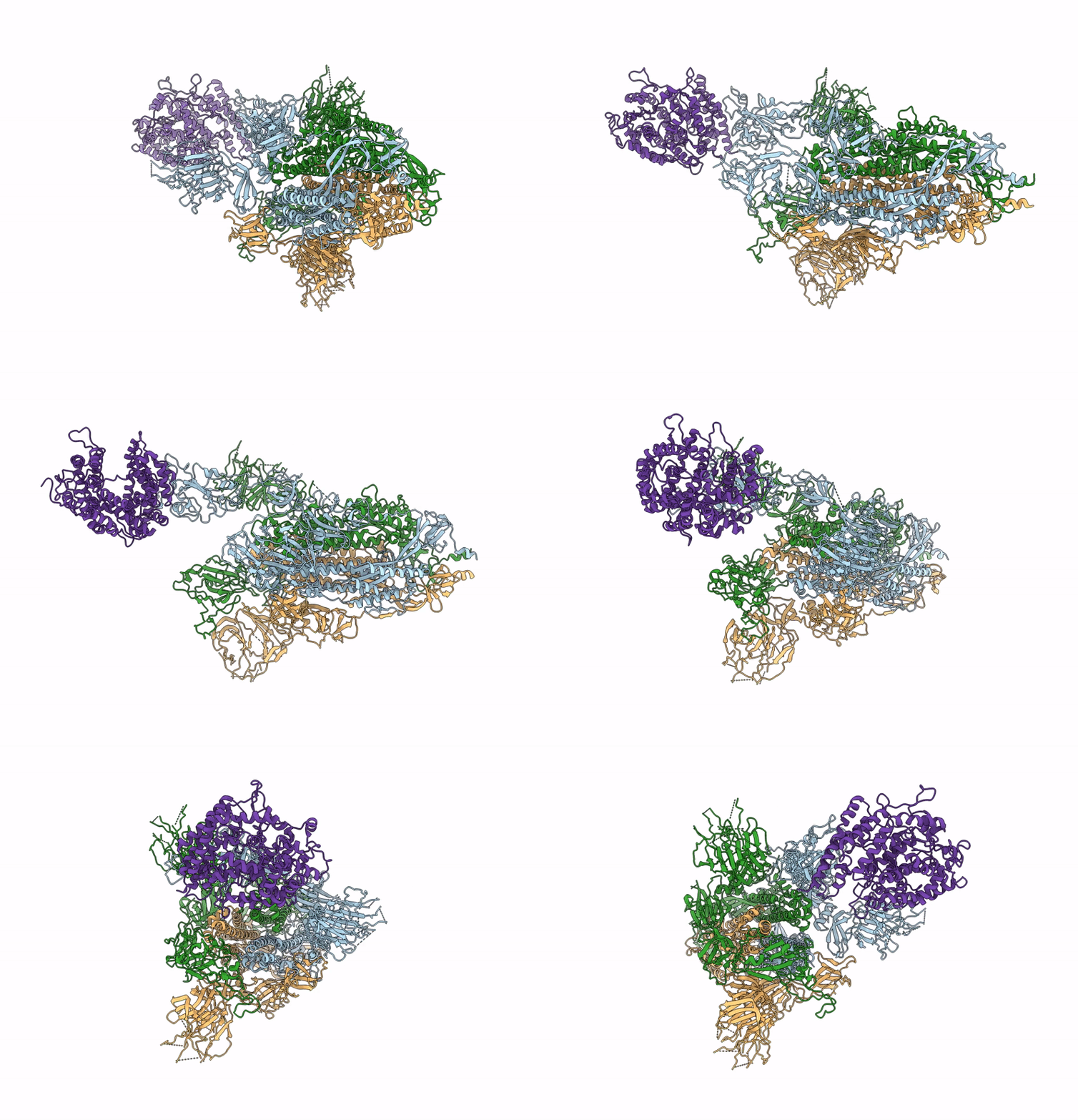
8TAZ image
Entry Detail
PDB ID:
8TAZ
EMDB ID:
Keywords:
Title:
Cryo-EM structure of mink variant Y453F trimeric spike protein bound to one mink ACE2 receptors
Biological Source:
PDB Version:
Deposition Date:
2023-06-28
Release Date:
2023-10-25
Method Details:
Experimental Method:
Resolution:
3.75 Å
Aggregation State:
PARTICLE
Reconstruction Method:
SINGLE PARTICLE
Macromolecular Entities
Polymer Type:polypeptide(L)
Description:Spike glycoprotein
Chain IDs:A, B, C
Chain Length:1269
Number of Molecules:3
Biological Source:Severe acute respiratory syndrome coronavirus 2
Polymer Type:polypeptide(L)
Description:Angiotensin-converting enzyme
Chain IDs:D
Chain Length:771
Number of Molecules:1
Biological Source:Neogale vison
Ligand Molecules
Primary Citation
Structural basis of the American mink ACE2 binding by Y453F trimeric spike glycoproteins of SARS-CoV-2.
J Med Virol 95 e29163 e29163 (2023)
PMID: 37842796 DOI: 10.1002/jmv.29163

Abstact

Severe Acute Respiratory Syndrome Coronavirus 2 (SARS-CoV-2) enters the host cell by binding to angiotensin-converting enzyme 2 (ACE2). While evolutionarily conserved, ACE2 receptors differ across various species and differential interactions with Spike (S) glycoproteins of SARS-CoV-2 viruses impact species specificity. Reverse zoonoses led to SARS-CoV-2 outbreaks on multiple American mink (Mustela vison) farms during the pandemic and gave rise to mink-associated S substitutions known for transmissibility between mink and zoonotic transmission to humans. In this study, we used bio-layer interferometry (BLI) to discern the differences in binding affinity between multiple human and mink-derived S glycoproteins of SARS-CoV-2 and their respective ACE2 receptors. Further, we conducted a structural analysis of a mink variant S glycoprotein and American mink ACE2 (mvACE2) using cryo-electron microscopy (cryo-EM), revealing four distinct conformations. We discovered a novel intermediary conformation where the mvACE2 receptor is bound to the receptor-binding domain (RBD) of the S glycoprotein in a "down" position, approximately 34° lower than previously reported "up" RBD. Finally, we compared residue interactions in the S-ACE2 complex interface of S glycoprotein conformations with varying RBD orientations. These findings provide valuable insights into the molecular mechanisms of SARS-CoV-2 entry.

Legend

Protein

Chemical

Disease

Primary Citation of related structures