8TAA image
Deposition Date 2023-06-27
Release Date 2024-07-03
Last Version Date 2025-02-05
Entry Detail
PDB ID:
8TAA
Keywords:
Title:
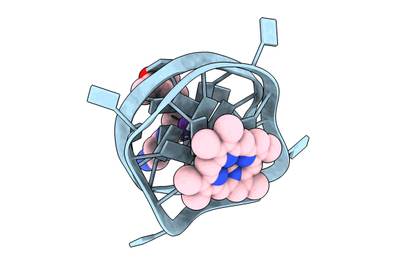
Right-left hybrid parallel G-quadruplex in complex with N-methyl mesoporphyrin
Biological Source:
Source Organism:
Homo sapiens (Taxon ID: 9606)
Method Details:
Experimental Method:
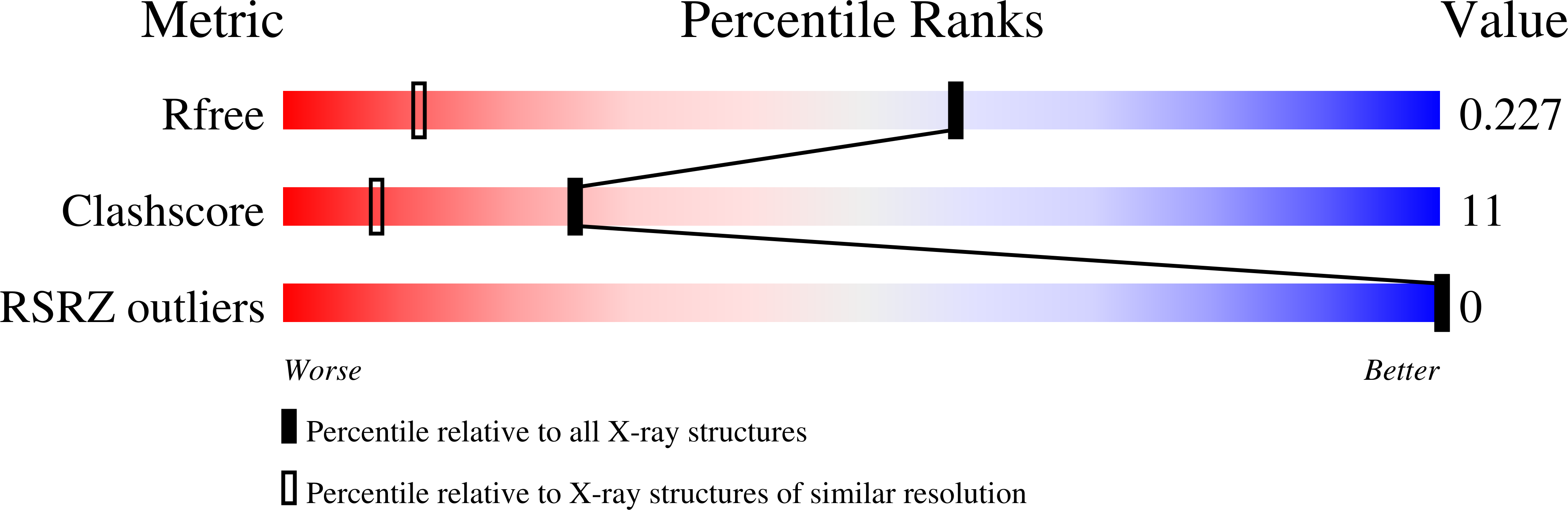
Resolution:
1.45 Å
R-Value Free:
0.22
R-Value Work:
0.17
R-Value Observed:
0.18
Space Group:
P 41 21 2
Macromolecular Entities
Polymer Type:polydeoxyribonucleotide
Molecule:DNA (25-MER)
Chain IDs:A
Chain Length:25
Number of Molecules:1
Biological Source:Homo sapiens
Primary Citation
Interaction of N-methylmesoporphyrin IX with a hybrid left-/right-handed G-quadruplex motif from the promoter of the SLC2A1 gene.
Nucleic Acids Res. 53 ? ? (2025)
PMID: 39704129 DOI: 10.1093/nar/gkae1208

Abstact

Left-handed G-quadruplexes (LHG4s) belong to a class of recently discovered noncanonical DNA structures under the larger umbrella of G-quadruplex DNAs (G4s). The biological relevance of these structures and their ability to be targeted with classical G4 ligands is underexplored. Here, we explore whether the putative LHG4 DNA sequence from the SLC2A1 oncogene promoter maintains its left-handed characteristics upon addition of nucleotides in the 5'- and 3'-direction from its genomic context. We also investigate whether this sequence interacts with a well-established G4 binder, N-methylmesoporphyrin IX (NMM). We employed biophysical and X-ray structural studies to address these questions. Our results indicate that the sequence d[G(TGG)3TGA(TGG)4] (termed here as SLC) adopts a two-subunit, four-tetrad hybrid left-/right-handed G4 (LH/RHG4) topology. Addition of 5'-G or 5'-GG abolishes the left-handed fold in one subunit, while the addition of 3'-C or 3'-CA maintains the original fold. X-ray crystal structure analyses show that SLC maintains the same hybrid LH/RHG4 fold in the solid state and that NMM stacks onto the right-handed subunit of SLC. NMM binds to SLC with a 1:1 stoichiometry and a moderate-to-tight binding constant of 15 μM-1. This work deepens our understanding of LHG4 structures and their binding with traditional G4 ligands.

Legend

Protein

Chemical

Disease

Primary Citation of related structures