8T9O image
Entry Detail
PDB ID:
8T9O
Keywords:
Title:
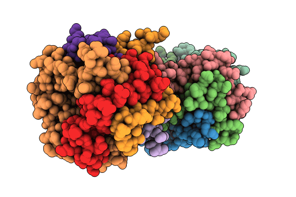
Crystal structure of CF, a heterohexamer of the 4-oxalocrotonate tautomerase (4-OT) family
Biological Source:
Source Organism:
Host Organism:
PDB Version:
Deposition Date:
2023-06-24
Release Date:
2023-12-06
Method Details:
Experimental Method:
Resolution:
2.70 Å
R-Value Free:
0.26
R-Value Work:
0.21
R-Value Observed:
0.21
Space Group:
C 1 2 1
Macromolecular Entities
Polymer Type:polypeptide(L)
Description:Tautomerase beta subunit
Chain IDs:A, E, F, G, H, L
Chain Length:71
Number of Molecules:6
Biological Source:Herbaspirillum
Polymer Type:polypeptide(L)
Description:Tautomerase alpha subunit
Chain IDs:B, C, D, I, J, K
Chain Length:67
Number of Molecules:6
Biological Source:Herbaspirillum
Primary Citation
Introduction of Asymmetry in the Fused 4-Oxalocrotonate Tautomerases.
Biochemistry 62 2461 2471 (2023)
PMID: 37490761 DOI: 10.1021/acs.biochem.3c00180

Abstact

Members of the 4-oxalocrotonate tautomerase (4-OT) subgroup in the tautomerase superfamily (TSF) are constructed from a single β-α-β unit and form homo- or heterohexamers, whereas those of the other four subgroups are composed of two consecutively joined β-α-β units and form trimers. A subset of sequences, double the length of the short 4-OTs, is found in the 4-OT subgroup. These "fused" 4-OTs form a separate subgroup that connects to the short 4-OTs in a sequence similarity network (SSN). The fused gene can be a template for the other four subgroups, resulting in the diversification of activity. Analysis of the SSN shows that multiple nodes in the fused 4-OTs connect to five linker nodes, which in turn connect to the short 4-OTs. Some fused 4-OTs are symmetric trimers and others are asymmetric trimers. The origin of this asymmetry was investigated by subjecting the sequences in three linker nodes and a closely associated fourth node to kinetic, mutagenic, and structural analyses. The results show that each sequence corresponds to the α- or β-subunit of a heterohexamer that functions as a 4-OT. Mutagenesis indicates that the key residues in both are αPro1 and βArg-11, like that of a typical 4-OT. Crystallographic analysis shows that both heterohexamers are asymmetric, where one heterodimer is flipped 180° relative to the other two heterodimers. The fusion of two subunits (α and β) of one asymmetric heterohexamer generates an asymmetric trimer with 4-OT activity. Hence, asymmetry can be introduced at the heterohexamer level and then retained in the fused trimers.

Legend

Protein

Chemical

Disease

Primary Citation of related structures