8T7D image
Entry Detail
PDB ID:
8T7D
Keywords:
Title:
Crystal structure of wild type IDH1 bound to compound 1
Biological Source:
Source Organism:
Host Organism:
PDB Version:
Deposition Date:
2023-06-20
Release Date:
2024-08-28
Method Details:
Experimental Method:
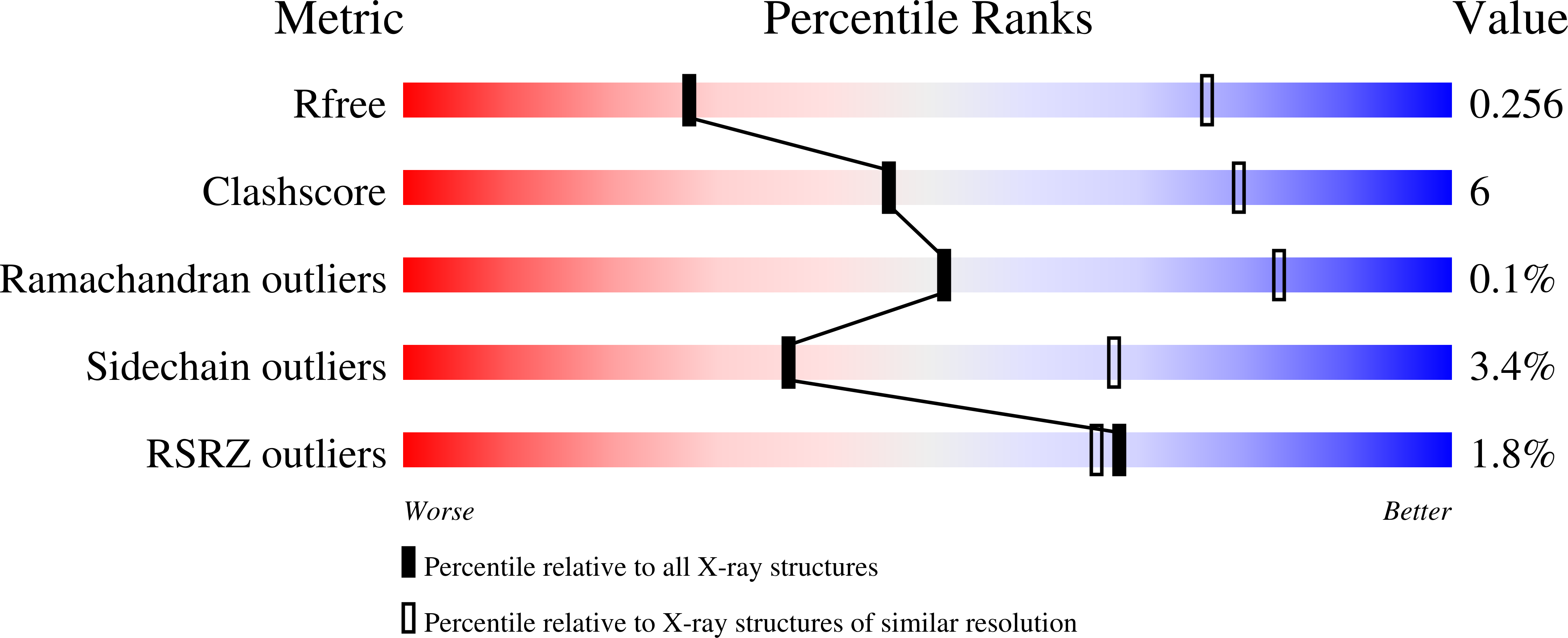
Resolution:
3.44 Å
R-Value Free:
0.26
R-Value Work:
0.24
R-Value Observed:
0.24
Space Group:
C 1 2 1
Macromolecular Entities
Polymer Type:polypeptide(L)
Description:Isocitrate dehydrogenase [NADP] cytoplasmic
Chain IDs:A, B, C, D
Chain Length:424
Number of Molecules:4
Biological Source:Homo sapiens
Ligand Molecules
Primary Citation
Biostructural, biochemical and biophysical studies of mutant IDH1.
Nat Commun 15 7877 7877 (2024)
PMID: 39251618 DOI: 10.1038/s41467-024-51692-0

Abstact

We report bio-structural, bio-chemical and bio-physical evidence demonstrating how small molecules can bind to both wild-type and mutant IDH1, but only inhibit the enzymatic activity of the mutant isoform. Enabled through x-ray crystallography, we characterized a series of small molecule inhibitors that bound to mutant IDH1 differently than the marketed inhibitor Ivosidenib, for which we have determined the x-ray crystal structure. Across the industry several mutant IDH1 inhibitor chemotypes bind to this allosteric IDH1 pocket and selectively inhibit the mutant enzyme. Detailed characterization by a variety of biophysical techniques and NMR studies led us to propose how compounds binding in the allosteric IDH1 R132H pocket inhibit the production of 2-Hydroxy glutarate.

Legend

Protein

Chemical

Disease

Primary Citation of related structures