Deposition Date
2023-06-12
Release Date
2023-12-27
Last Version Date
2024-02-14
Entry Detail
PDB ID:
8T57
Keywords:
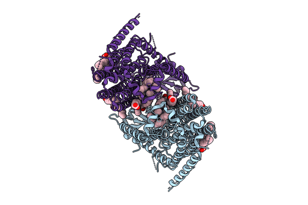
Title:
Structure of mechanically activated ion channel OSCA2.3 in peptidiscs
Biological Source:
Source Organism(s):
Arabidopsis thaliana (Taxon ID: 3702)
Expression System(s):
Method Details:
Experimental Method:
Resolution:
2.70 Å
Aggregation State:
PARTICLE
Reconstruction Method:
SINGLE PARTICLE