8T3O image
Entry Detail
PDB ID:
8T3O
EMDB ID:
Title:
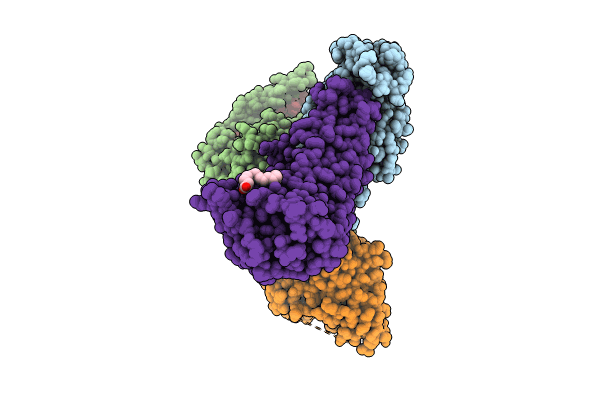
Cryo-EM structure of the TUG-891 bound FFA4-Gq complex
Biological Source:
Source Organism:
Host Organism:
PDB Version:
Deposition Date:
2023-06-07
Release Date:
2024-01-17
Method Details:
Experimental Method:
Resolution:
3.06 Å
Aggregation State:
PARTICLE
Reconstruction Method:
SINGLE PARTICLE
Macromolecular Entities
Polymer Type:polypeptide(L)
Description:Guanine nucleotide-binding protein G(q)
Chain IDs:D (auth: A)
Chain Length:230
Number of Molecules:1
Biological Source:Homo sapiens
Polymer Type:polypeptide(L)
Description:Guanine nucleotide-binding protein G(I)/G(S)/G(T) subunit beta-1
Chain IDs:B
Chain Length:342
Number of Molecules:1
Biological Source:Homo sapiens
Polymer Type:polypeptide(L)
Description:Guanine nucleotide-binding protein G(I)/G(S)/G(O) subunit gamma-2
Chain IDs:C (auth: G)
Chain Length:57
Number of Molecules:1
Biological Source:Homo sapiens
Polymer Type:polypeptide(L)
Description:scFv16
Chain IDs:E (auth: N)
Chain Length:266
Number of Molecules:1
Biological Source:Mus musculus
Polymer Type:polypeptide(L)
Description:Free fatty acid receptor 4
Chain IDs:A (auth: R)
Chain Length:297
Number of Molecules:1
Biological Source:Homo sapiens
Primary Citation
Structural basis for the ligand recognition and signaling of free fatty acid receptors.
Sci Adv 10 eadj2384 eadj2384 (2024)
PMID: 38198545 DOI: 10.1126/sciadv.adj2384

Abstact

Free fatty acid receptors 1 to 4 (FFA1 to FFA4) are class A G protein-coupled receptors (GPCRs). FFA1 to FFA3 share substantial sequence similarity, whereas FFA4 is unrelated. However, FFA1 and FFA4 are activated by long-chain fatty acids, while FFA2 and FFA3 respond to short-chain fatty acids generated by intestinal microbiota. FFA1, FFA2, and FFA4 are potential drug targets for metabolic and inflammatory conditions. Here, we determined the active structures of FFA1 and FFA4 bound to docosahexaenoic acid, FFA4 bound to the synthetic agonist TUG-891, and butyrate-bound FFA2, each complexed with an engineered heterotrimeric Gq protein (miniGq), by cryo-electron microscopy. Together with computational simulations and mutagenesis studies, we elucidated the similarities and differences in the binding modes of fatty acid ligands to their respective GPCRs. Our findings unveiled distinct mechanisms of receptor activation and G protein coupling. We anticipate that these outcomes will facilitate structure-based drug development and underpin future research on this group of GPCRs.

Legend

Protein

Chemical

Disease

Primary Citation of related structures