
Deposition Date
2023-06-06
Release Date
2024-06-12
Last Version Date
2024-10-23
Entry Detail
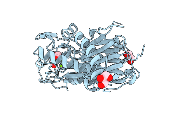
PDB ID:
8T2J
Keywords:
Title:
Structure of the catalytic domain of PPM1D/Wip1 serine/threonine phosphatase
Biological Source:
Source Organism(s):
Homo sapiens (Taxon ID: 9606)
Expression System(s):
Method Details:
Experimental Method:
Resolution:
1.80 Å
R-Value Free:
0.21
R-Value Work:
0.17
R-Value Observed:
0.17
Space Group:
P 1 21 1
