Deposition Date
2023-05-29
Release Date
2024-02-07
Last Version Date
2024-10-16
Entry Detail
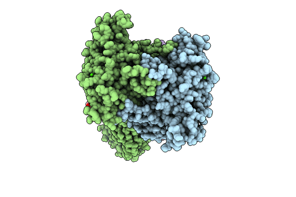
PDB ID:
8SZH
Keywords:
Title:
Cryo-EM structure of cinacalcet-bound human calcium-sensing receptor CaSR-Gi complex in lipid nanodiscs
Biological Source:
Source Organism(s):
Homo sapiens (Taxon ID: 9606)
Expression System(s):
Method Details:
Experimental Method:
Resolution:
3.10 Å
Aggregation State:
PARTICLE
Reconstruction Method:
SINGLE PARTICLE