8SYF image
Entry Detail
PDB ID:
8SYF
EMDB ID:
Keywords:
Title:
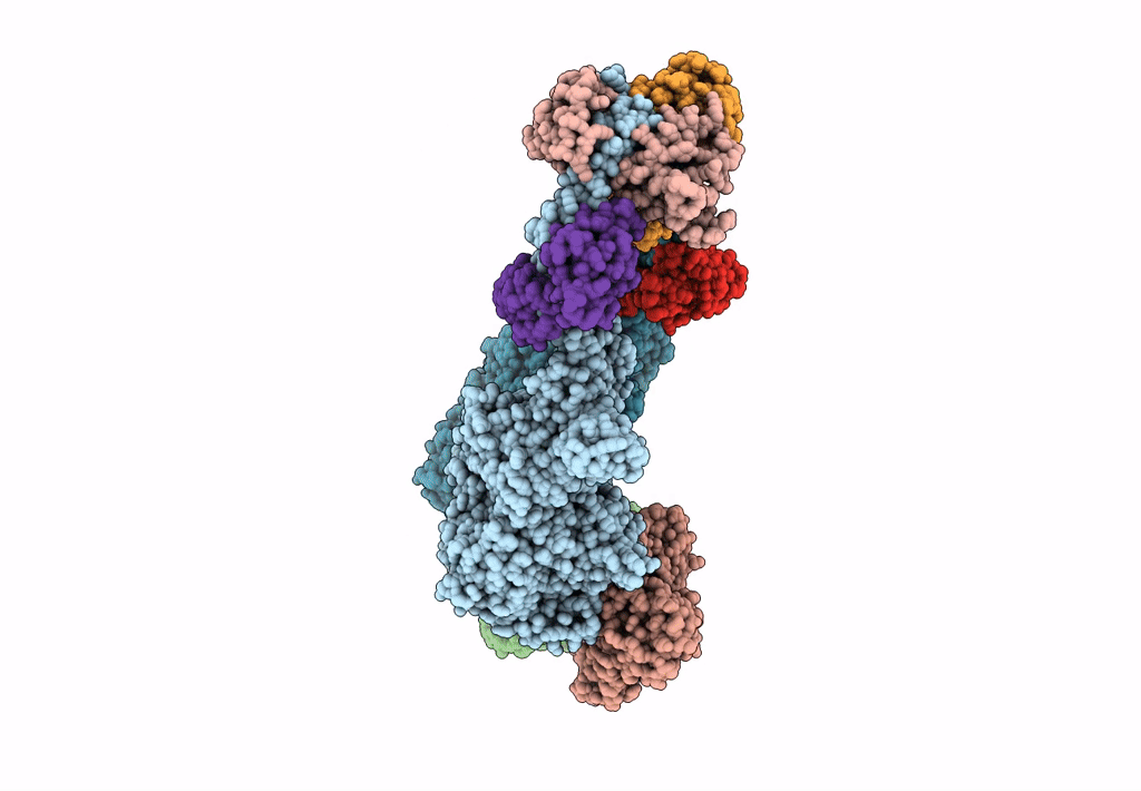
Homology model of Acto-HMM complex in ADP-state. Chicken smooth muscle HMM and chicken pectoralis actin
Biological Source:
Source Organism:
PDB Version:
Deposition Date:
2023-05-25
Release Date:
2023-08-30
Method Details:
Experimental Method:
Resolution:
19.00 Å
Aggregation State:
FILAMENT
Reconstruction Method:
SINGLE PARTICLE
Macromolecular Entities
Polymer Type:polypeptide(L)
Description:Myosin, heavy chain 11, smooth muscle
Chain IDs:A, B
Chain Length:843
Number of Molecules:2
Biological Source:Gallus gallus
Polymer Type:polypeptide(L)
Description:Actin, alpha skeletal muscle
Chain IDs:C, D
Chain Length:375
Number of Molecules:2
Biological Source:Gallus gallus
Polymer Type:polypeptide(L)
Description:Myosin light polypeptide 6
Chain IDs:E, H
Chain Length:148
Number of Molecules:2
Biological Source:Gallus gallus
Polymer Type:polypeptide(L)
Description:Myosin regulatory light chain 2, smooth muscle major isoform
Chain IDs:F, G
Chain Length:143
Number of Molecules:2
Biological Source:Gallus gallus
Ligand Molecules
Primary Citation
Double-headed binding of myosin II to F-actin shows the effect of strain on head structure.
J.Struct.Biol. 215 107995 107995 (2023)
PMID: 37414375 DOI: 10.1016/j.jsb.2023.107995

Abstact

Force production in muscle is achieved through the interaction of myosin and actin. Strong binding states in active muscle are associated with Mg·ADP bound to the active site; release of Mg·ADP allows rebinding of ATP and dissociation from actin. Thus, Mg·ADP binding is positioned for adaptation as a force sensor. Mechanical loads on the lever arm can affect the ability of myosin to release Mg·ADP but exactly how this is done is poorly defined. Here we use F-actin decorated with double-headed smooth muscle myosin fragments in the presence of Mg·ADP to visualize the effect of internally supplied tension on the paired lever arms using cryoEM. The interaction of the paired heads with two adjacent actin subunits is predicted to place one lever arm under positive and the other under negative strain. The converter domain is believed to be the most flexible domain within myosin head. Our results, instead, point to the segment of heavy chain between the essential and regulatory light chains as the location of the largest structural change. Moreover, our results suggest no large changes in the myosin coiled coil tail as the locus of strain relief when both heads bind F-actin. The method would be adaptable to double-headed members of the myosin family. We anticipate that the study of actin-myosin interaction using double-headed fragments enables visualization of domains that are typically noisy in decoration with single-headed fragments.

Legend

Protein

Chemical

Disease

Primary Citation of related structures