8SYD image
Entry Detail
PDB ID:
8SYD
Keywords:
Title:
X-ray crystal structure of UDP-2,3-diacetamido-2,3-dideoxy-glucuronic acid-2-epimerase from Thermus thermophilus strain HB27, D98N variant in the presence of UDP-2,3-diacetamido-2,3-dideoxy-glucuronic acid and UDP-N-acetylglucosamine at pH 6
Biological Source:
Source Organism:
PDB Version:
Deposition Date:
2023-05-25
Release Date:
2023-09-13
Method Details:
Experimental Method:
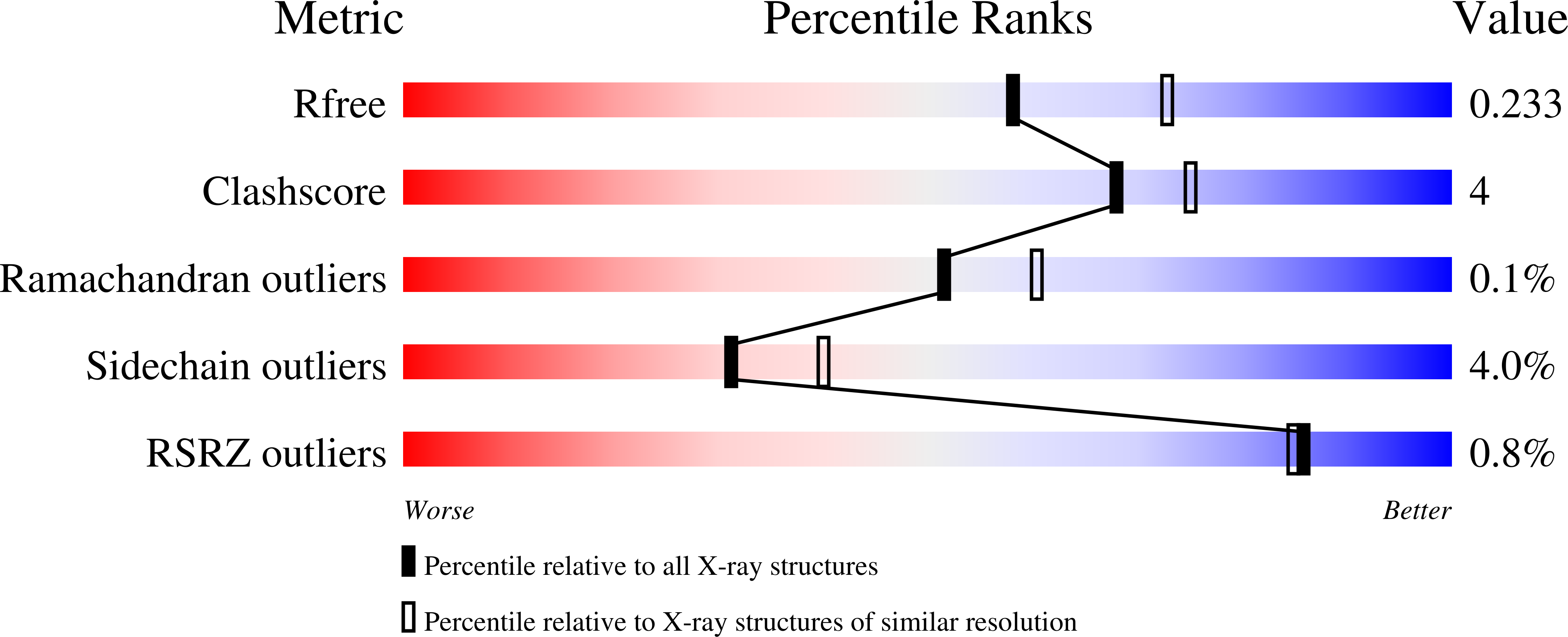
Resolution:
2.20 Å
R-Value Free:
0.22
R-Value Work:
0.17
R-Value Observed:
0.17
Space Group:
P 1 21 1
Macromolecular Entities
Polymer Type:polypeptide(L)
Description:UDP-2,3-diacetamido-2,3-dideoxy-glucuronic acid-2-epimerase
Mutations:D98N
Chain IDs:A, B
Chain Length:385
Number of Molecules:2
Biological Source:Thermus thermophilus HB27
Primary Citation
Structural analysis of a bacterial UDP-sugar 2-epimerase reveals the active site architecture before and after catalysis.
J.Biol.Chem. 299 105200 105200 (2023)
PMID: 37660908 DOI: 10.1016/j.jbc.2023.105200

Abstact

The sugar, 2,3-diacetamido-2,3-dideoxy-d-mannuronic acid, was first identified ∼40 years ago in the O-antigen of Pseudomonas aeruginosa O:3,a,d. Since then, it has been observed on the O-antigens of various pathogenic Gram-negative bacteria including Bordetella pertussis, Escherichia albertii, and Pseudomonas mediterranea. Previous studies have established that five enzymes are required for its biosynthesis beginning with uridine dinucleotide (UDP)-N-acetyl-d-glucosamine (UDP-GlcNAc). The final step in the pathway is catalyzed by a 2-epimerase, which utilizes UDP-2,3-diacetamido-2,3-dideoxy-d-glucuronic acid as its substrate. Curious as to whether this biochemical pathway is found in extreme thermophiles, we examined the published genome sequence for Thermus thermophilus HB27 and identified five ORFs that could possibly encode for the required enzymes. The focus of this investigation is on the ORF WP_011172736, which we demonstrate encodes for a 2-epimerase. For this investigation, ten high resolution X-ray crystallographic structures were determined to resolutions of 2.3 Å or higher. The models have revealed the manner in which the 2-epimerase anchors its UDP-sugar substrate as well as its UDP-sugar product into the active site. In addition, this study reveals for the first time the manner in which any sugar 2-epimerase can simultaneously bind UDP-sugars in both the active site and the allosteric binding region. We have also demonstrated that the T. thermophilus enzyme is allosterically regulated by UDP-GlcNAc. Whereas the sugar 2-epimerases that function on UDP-GlcNAc have been the focus of past biochemical and structural analyses, this is the first detailed investigation of a 2-epimerase that specifically utilizes UDP-2,3-diacetamido-2,3-dideoxy-d-glucuronic acid as its substrate.

Legend

Protein

Chemical

Disease

Primary Citation of related structures