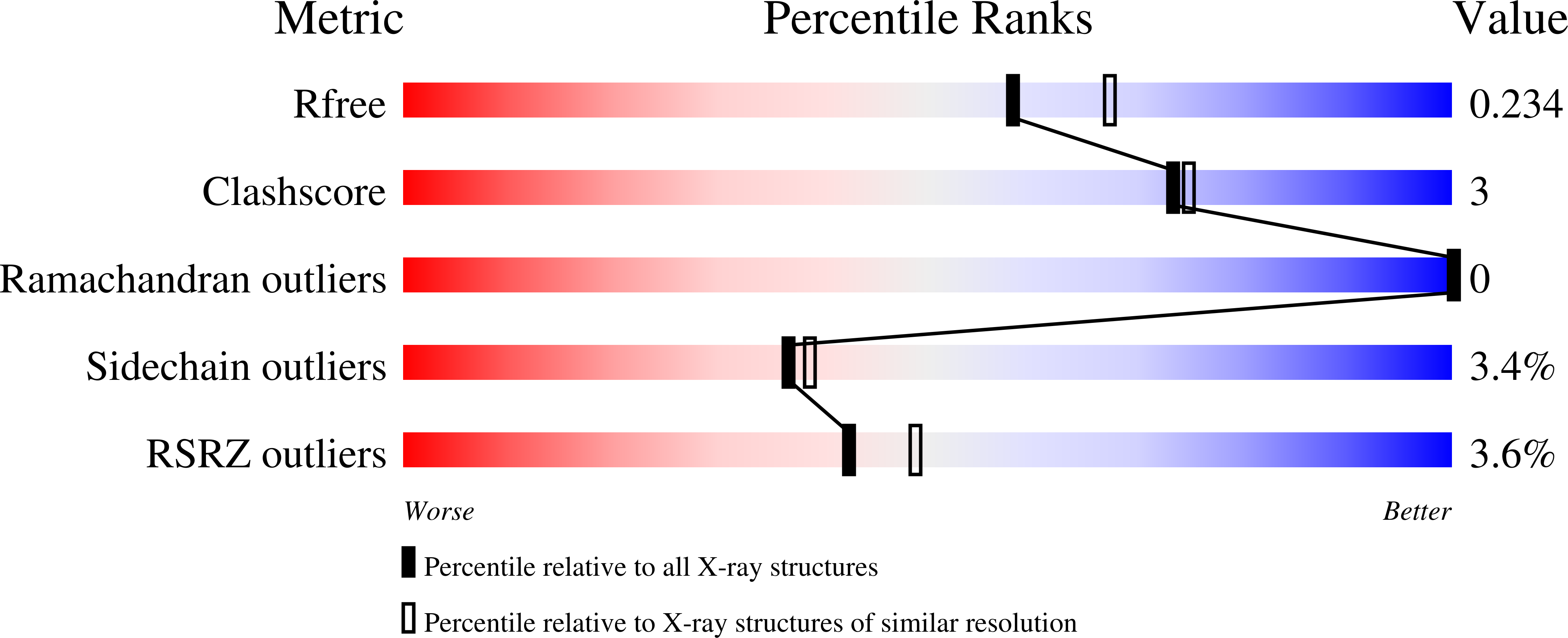
8SWP image
Entry Detail
PDB ID:
8SWP
Keywords:
Title:
Structure of K. lactis PNP bound to hypoxanthine
Biological Source:
PDB Version:
Deposition Date:
2023-05-19
Release Date:
2023-10-18
Method Details:
Experimental Method:
Resolution:
2.10 Å
R-Value Free:
0.23
R-Value Work:
0.19
R-Value Observed:
0.20
Space Group:
P 1 21 1
Macromolecular Entities
Polymer Type:polypeptide(L)
Description:Purine nucleoside phosphorylase
Chain IDs:A, B, C, D, E, F
Chain Length:307
Number of Molecules:6
Biological Source:Kluyveromyces lactis NRRL Y-1140
Modified Residue
Compound ID Chain ID Parent Comp ID Details 2D Image
MSE A MET modified residue
Primary Citation
Phosphate Binding in PNP Alters Transition-State Analogue Affinity and Subunit Cooperativity.
Biochemistry 62 3116 3125 (2023)
PMID: 37812583 DOI: 10.1021/acs.biochem.3c00264

Abstact

Purine nucleoside phosphorylases (PNPs) catalyze the phosphorolysis of 6-oxypurine nucleosides with an HPO42- dianion nucleophile. Nucleosides and phosphate occupy distinct pockets in the PNP active site. Evaluation of the HPO42- site by mutagenesis, cooperative binding studies, and thermodynamic and structural analysis demonstrate that alterations in the HPO42- binding site can render PNP inactive and significantly impact subunit cooperativity and binding to transition-state analogue inhibitors. Cooperative interactions between the cationic transition-state analogue and the anionic HPO42- nucleophile demonstrate the importance of reforming the transition-state ensemble for optimal inhibition with transition-state analogues. Altered phosphate binding in the catalytic site mutants helps to explain one of the known lethal PNP deficiency syndromes in humans.

Legend

Protein

Chemical

Disease

Primary Citation of related structures