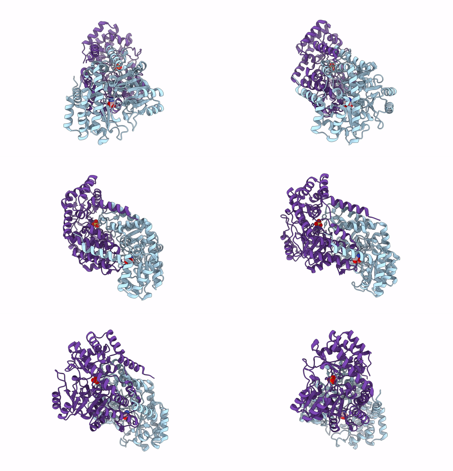
8SSY image
Entry Detail
PDB ID:
8SSY
Keywords:
Title:
Room-temperature X-ray structure of Thermus thermophilus serine hydroxymethyltransferase (SHMT) bound with D-Ser in a pseudo-Michaelis complex
Biological Source:
Source Organism:
Host Organism:
PDB Version:
Deposition Date:
2023-05-09
Release Date:
2023-08-16
Method Details:
Experimental Method:
Resolution:
1.80 Å
R-Value Free:
0.16
R-Value Work:
0.13
R-Value Observed:
0.13
Space Group:
P 1 21 1
Macromolecular Entities
Polymer Type:polypeptide(L)
Description:Serine hydroxymethyltransferase
Chain IDs:A, B
Chain Length:405
Number of Molecules:2
Biological Source:Thermus thermophilus HB8
Modified Residue
Compound ID Chain ID Parent Comp ID Details 2D Image
LLP A LYS modified residue
Primary Citation
Revealing protonation states and tracking substrate in serine hydroxymethyltransferase with room-temperature X-ray and neutron crystallography.
Commun Chem 6 162 162 (2023)
PMID: 37532884 DOI: 10.1038/s42004-023-00964-9

Abstact

Pyridoxal 5'-phosphate (PLP)-dependent enzymes utilize a vitamin B6-derived cofactor to perform a myriad of chemical transformations on amino acids and other small molecules. Some PLP-dependent enzymes, such as serine hydroxymethyltransferase (SHMT), are promising drug targets for the design of small-molecule antimicrobials and anticancer therapeutics, while others have been used to synthesize pharmaceutical building blocks. Understanding PLP-dependent catalysis and the reaction specificity is crucial to advance structure-assisted drug design and enzyme engineering. Here we report the direct determination of the protonation states in the active site of Thermus thermophilus SHMT (TthSHMT) in the internal aldimine state using room-temperature joint X-ray/neutron crystallography. Conserved active site architecture of the model enzyme TthSHMT and of human mitochondrial SHMT (hSHMT2) were compared by obtaining a room-temperature X-ray structure of hSHMT2, suggesting identical protonation states in the human enzyme. The amino acid substrate serine pathway through the TthSHMT active site cavity was tracked, revealing the peripheral and cationic binding sites that correspond to the pre-Michaelis and pseudo-Michaelis complexes, respectively. At the peripheral binding site, the substrate is bound in the zwitterionic form. By analyzing the observed protonation states, Glu53, but not His residues, is proposed as the general base catalyst, orchestrating the retro-aldol transformation of L-serine into glycine.

Legend

Protein

Chemical

Disease

Primary Citation of related structures