8SMO image
Deposition Date 2023-04-26
Release Date 2024-05-01
Last Version Date 2025-05-14
Entry Detail
PDB ID:
8SMO
Title:
Crystal structure of the complex between truncated MLLE domain of PABPC1 and engineered superPAM2 peptide
Biological Source:
Source Organism:
Host Organism:
Method Details:
Experimental Method:
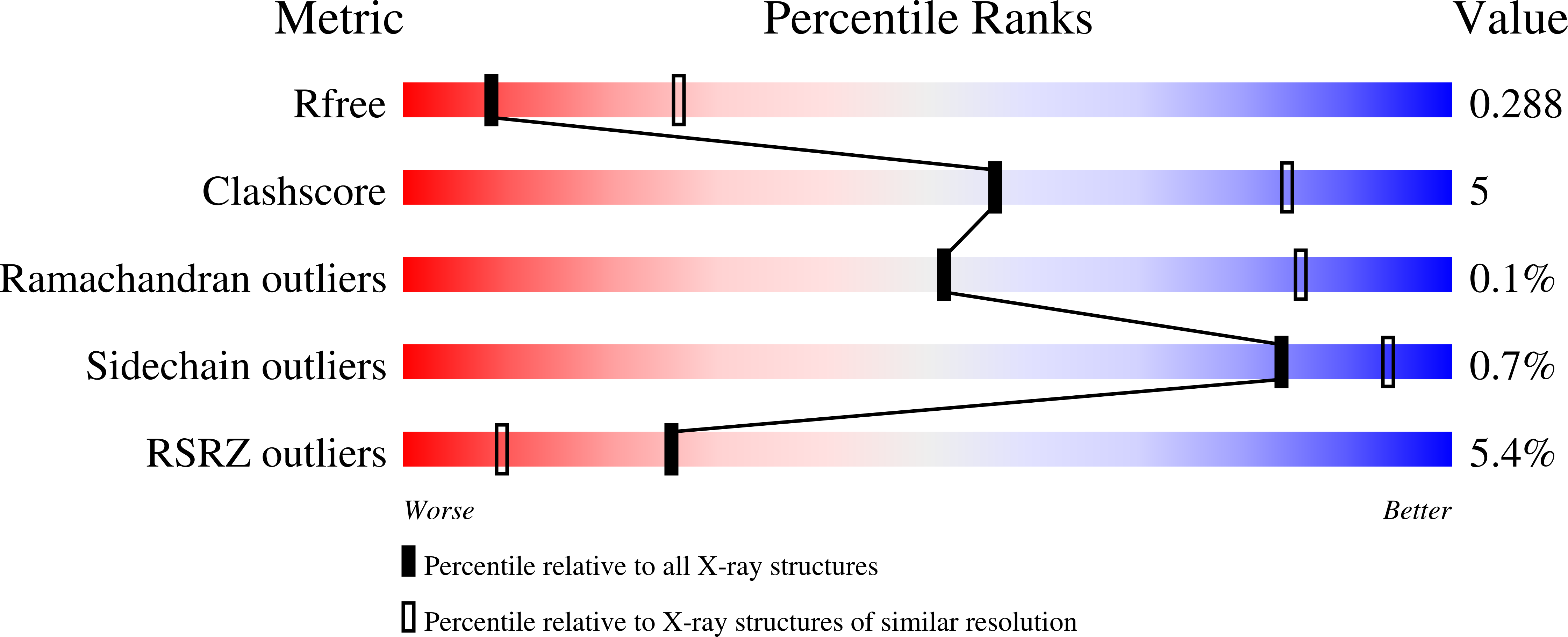
Resolution:
3.00 Å
R-Value Free:
0.29
R-Value Work:
0.25
R-Value Observed:
0.25
Space Group:
P 31
Macromolecular Entities
Polymer Type:polypeptide(L)
Molecule:Polyadenylate-binding protein 1
Gene (Uniprot):PABPC1
Chain IDs:A, C, E, G, I, K, M, O
Chain Length:76
Number of Molecules:8
Biological Source:Homo sapiens
Polymer Type:polypeptide(L)
Molecule:superPAM2
Chain IDs:B, D, F, H, J, L, N, P
Chain Length:22
Number of Molecules:8
Biological Source:synthetic construct
Ligand Molecules
Primary Citation
Deep Mutational Scanning of an Engineered High-affinity Ligand of the poly(A) Binding Protein MLLE Domain.
J.Mol.Biol. 437 169120 169120 (2025)
PMID: 40180125 DOI: 10.1016/j.jmb.2025.169120

Abstact

The MLLE domain is a peptide-binding domain found in the poly(A) binding protein (PABP) and the ubiquitin protein E3 ligase N-recognin 5 (UBR5) that recognizes a conserved motif, named PABP-interacting motif 2 (PAM2). The majority of PAM2 sequences bind to MLLE domains with low-micromolar affinity. Here, we designed a chimeric PAM2 peptide termed super PAM2 (sPAM2) by combining classical and trinucleotide repeat-containing 6 (TNRC6)-like binding modes to create a superior binder for the MLLE domain. The crystal structure of the PABPC1 MLLE-sPAM2 complex shows a crucial role of conserved sPAM2 leucine, phenylalanine and tryptophan residues in the interaction. We used deep mutational scanning (DMS) coupled with isothermal titration calorimetry (ITC) to characterize the specificity profiles for PABPC1 and UBR5 MLLE. The best sPAM2 sequence binds to PABPC1 MLLE with low-nanomolar affinity and nearly 20-fold more tightly than the best natural PAM2 sequence. This suggests that the affinities of natural PAM2 sequences are tuned to control their binding to PABPC1 and UBR5. Our study will aid in the discovery of new PAM2-containing proteins (PACs) and facilitate in vivo studies of PAM2-mediated cellular pathways.

Legend

Protein

Chemical

Disease

Primary Citation of related structures