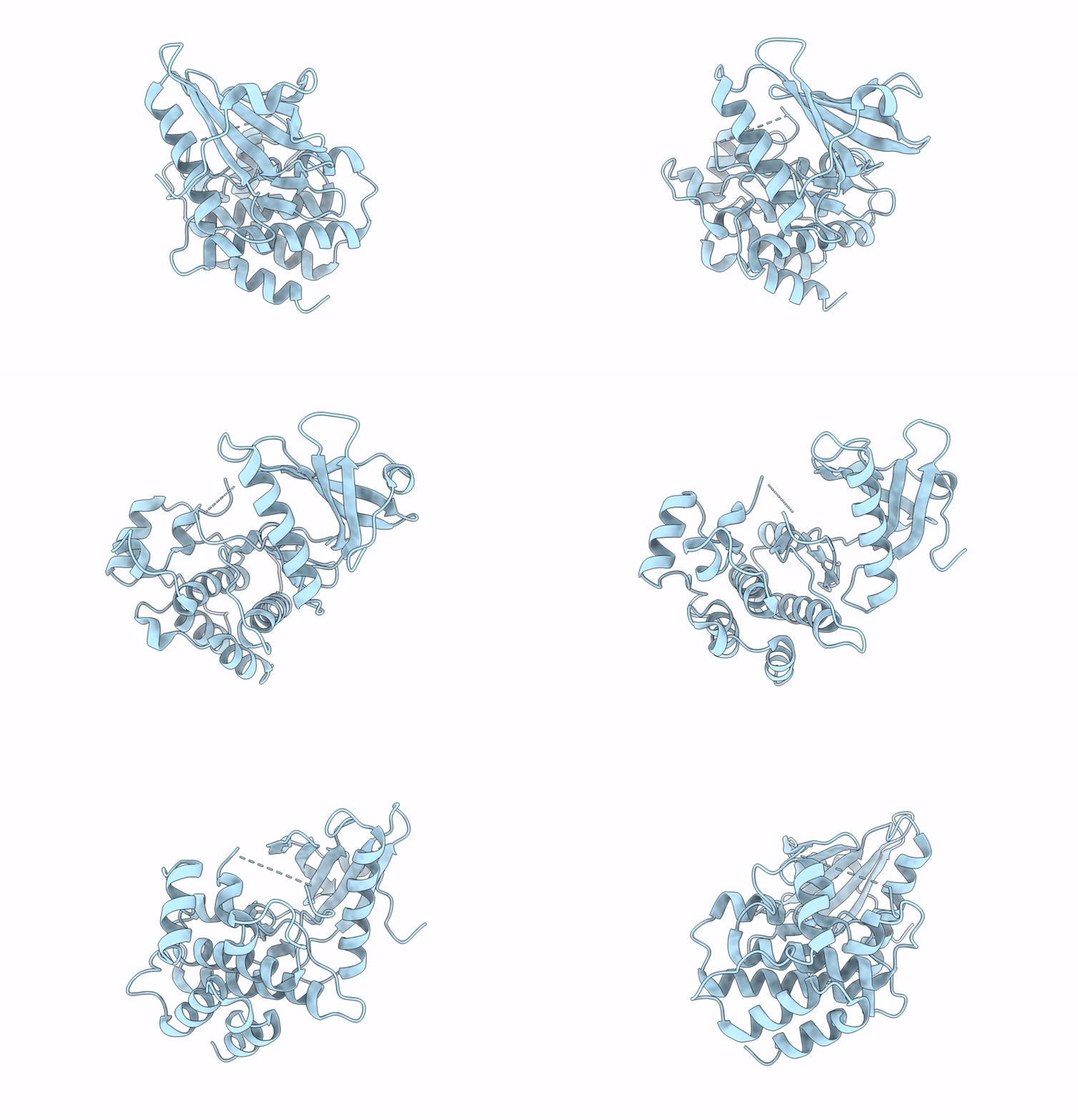
8SLZ image
Deposition Date 2023-04-25
Release Date 2023-11-08
Last Version Date 2024-11-06
Entry Detail
PDB ID:
8SLZ
Keywords:
Title:
Crystal structure of phosphorylated (T357/S358) human MLKL pseudokinase domain
Biological Source:
Source Organism:
Homo sapiens (Taxon ID: 9606)
Host Organism:
Method Details:
Experimental Method:
Resolution:
2.30 Å
R-Value Free:
0.23
R-Value Work:
0.20
R-Value Observed:
0.20
Space Group:
C 2 2 21
Macromolecular Entities
Polymer Type:polypeptide(L)
Molecule:Mixed lineage kinase domain-like protein
Gene (Uniprot):MLKL
Chain IDs:A
Chain Length:287
Number of Molecules:1
Biological Source:Homo sapiens
Primary Citation
Phosphorylation-dependent pseudokinase domain dimerization drives full-length MLKL oligomerization.
Nat Commun 14 6804 6804 (2023)
PMID: 37884510 DOI: 10.1038/s41467-023-42255-w

Abstact

The necroptosis pathway is a lytic, pro-inflammatory mode of cell death that is widely implicated in human disease, including renal, pulmonary, gut and skin inflammatory pathologies. The precise mechanism of the terminal steps in the pathway, where the RIPK3 kinase phosphorylates and triggers a conformation change and oligomerization of the terminal pathway effector, MLKL, are only emerging. Here, we structurally identify RIPK3-mediated phosphorylation of the human MLKL activation loop as a cue for MLKL pseudokinase domain dimerization. MLKL pseudokinase domain dimerization subsequently drives formation of elongated homotetramers. Negative stain electron microscopy and modelling support nucleation of the MLKL tetramer assembly by a central coiled coil formed by the extended, ~80 Å brace helix that connects the pseudokinase and executioner four-helix bundle domains. Mutational data assert MLKL tetramerization as an essential prerequisite step to enable the release and reorganization of four-helix bundle domains for membrane permeabilization and cell death.

Legend

Protein

Chemical

Disease

Primary Citation of related structures