Deposition Date
2023-04-21
Release Date
2023-06-07
Last Version Date
2024-10-23
Entry Detail
PDB ID:
8SLB
Keywords:
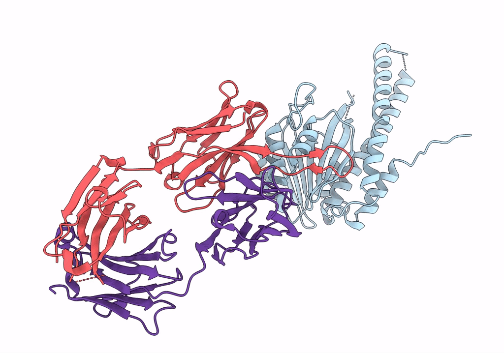
Title:
X-ray structure of CorA N-terminal domain in complex with conformation-specific synthetic antibody C12
Biological Source:
Source Organism(s):
Thermotoga maritima MSB8 (Taxon ID: 243274)
Homo sapiens (Taxon ID: 9606)
Homo sapiens (Taxon ID: 9606)
Expression System(s):
Method Details:
Experimental Method:
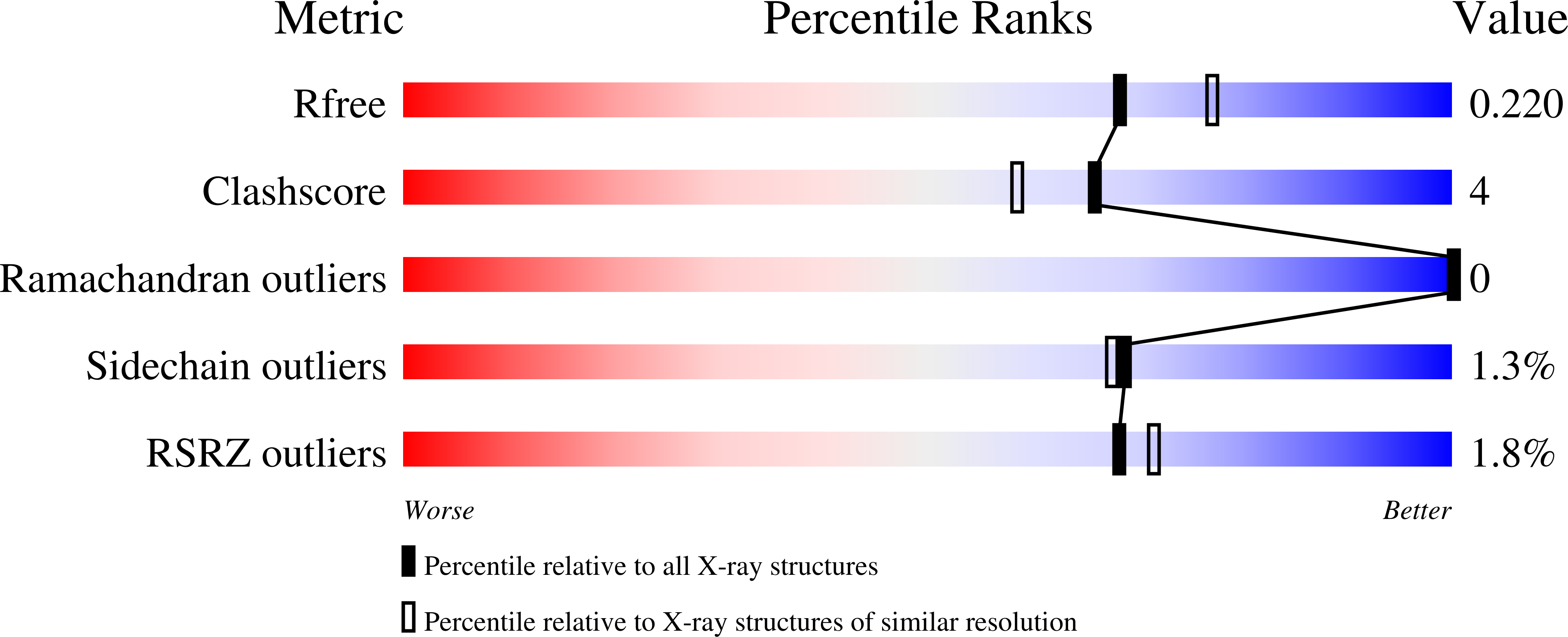
Resolution:
2.04 Å
R-Value Free:
0.21
R-Value Work:
0.16
R-Value Observed:
0.16
Space Group:
I 1 2 1