8SJ4 image
Entry Detail
PDB ID:
8SJ4
Keywords:
Title:
8F3-1H9-Ara h 6
Biological Source:
Source Organism:
Host Organism:
PDB Version:
Deposition Date:
2023-04-17
Release Date:
2024-12-25
Method Details:
Experimental Method:
Resolution:
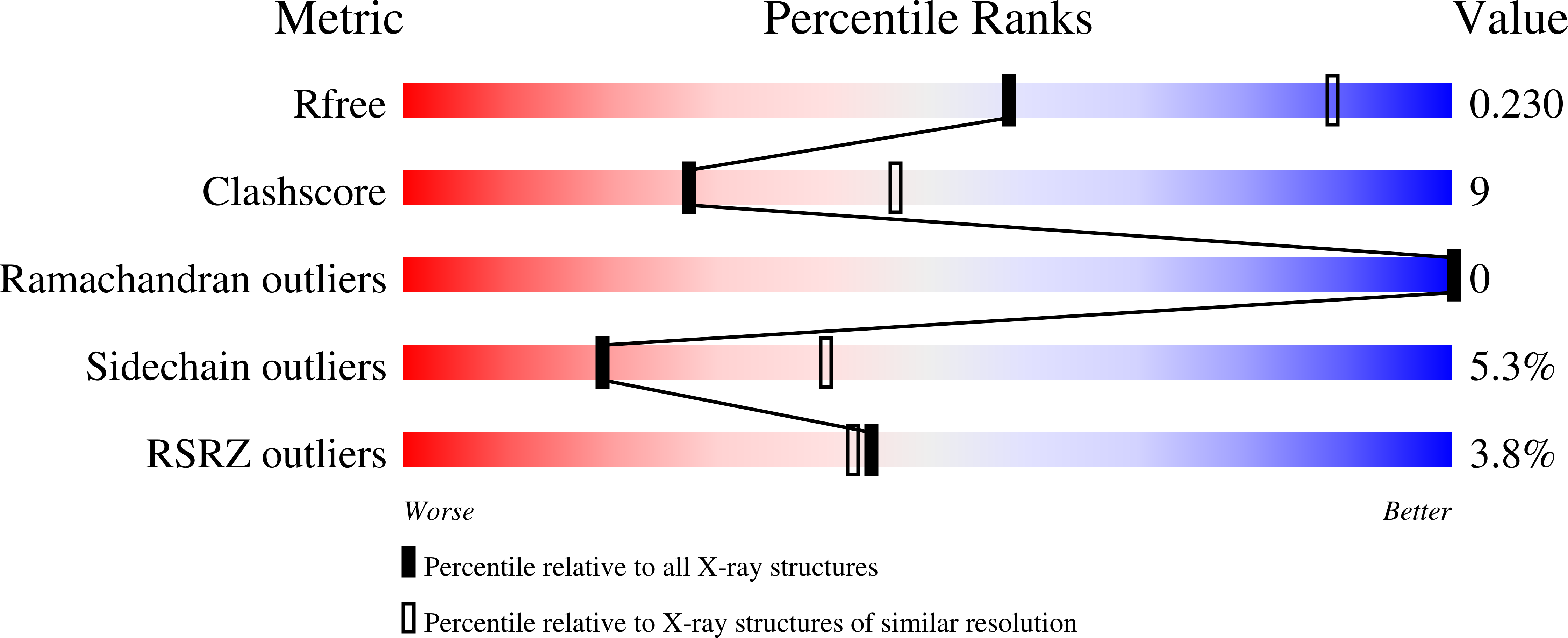
2.67 Å
R-Value Free:
0.23
R-Value Work:
0.19
R-Value Observed:
0.19
Space Group:
P 1 21 1
Macromolecular Entities
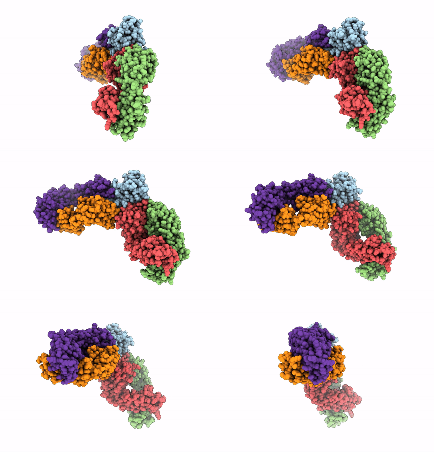
Polymer Type:polypeptide(L)
Description:Conglutin
Chain IDs:E (auth: A)
Chain Length:122
Number of Molecules:1
Biological Source:Arachis hypogaea
Polymer Type:polypeptide(L)
Description:8F3 light chain kappa
Chain IDs:C (auth: D)
Chain Length:214
Number of Molecules:1
Biological Source:Homo sapiens
Polymer Type:polypeptide(L)
Description:8F3 heavy chain
Chain IDs:D (auth: F)
Chain Length:225
Number of Molecules:1
Biological Source:Homo sapiens
Polymer Type:polypeptide(L)
Description:1H9 heavy chain
Chain IDs:A (auth: H)
Chain Length:218
Number of Molecules:1
Biological Source:Homo sapiens
Polymer Type:polypeptide(L)
Description:1H9 light chain kappa
Chain IDs:B (auth: L)
Chain Length:214
Number of Molecules:1
Biological Source:Homo sapiens
Primary Citation
Structural determinants of peanut induced anaphylaxis.
J.Allergy Clin.Immunol. ? ? ? (2025)
PMID: 39805366 DOI: 10.1016/j.jaci.2024.12.1095

Abstact

BACKGROUND Human IgE mAbs recognizing peanut allergens have recently become available, but we lack a detailed understanding of how these IgEs target allergens. OBJECTIVE We sought to determine the molecular details of the antibody-allergen interaction for a panel of clinically important human IgE mAbs and to develop strategies to disrupt disease causing antibody-allergen interactions. METHODS We identified candidates from a panel of epitope binned human IgE mAbs that recognize 2 important and homologous peanut allergens, Ara h 2 and Ara h 6. Crystal structures were determined revealing the interfaces (antigenic sites) of exemplars of 5 common IgE bins. RESULTS Among the common antigenic sites on Ara h 2 and Ara h 6, 2 sites (A and B) are highly conserved between the allergens, explaining the cross-reactivity of antibodies that recognize these sites. Three sites (C, D, and F) involve residues that are not conserved between the allergens. Of the 5 common sites, 3 sites (B, C, and D) involve residues that are near each other only when the allergens are properly folded, such that these sites are conformational. Two additional sites (sites A and F) involve largely linear stretches of amino acids. Site F targeted antibody, 38B7, binds to a peptide sequence DPYSPOHS, in which hydroxylation of the last proline is critical for binding. This sequence is repeated 2 or 3 times depending on the Ara h 2 isoform, enabling 38B7 to induce anaphylaxis as a single mAb, without a second antibody. We have mutated key residues in each site and created a panel of hypoallergens, having reduced IgE mAb binding and lacking the ability to induce anaphylaxis in our murine model. CONCLUSION We created a structural map of the IgE antibody response to the most important peanut allergen proteins to enable the design of new allergy immunotherapies and vaccines.

Legend

Protein

Chemical

Disease

Primary Citation of related structures