Deposition Date
2023-04-03
Release Date
2023-11-08
Last Version Date
2023-11-29
Entry Detail
PDB ID:
8SBM
Keywords:
Title:
Crystal structure of the wild-type Catalytic ATP-binding domain of Mtb DosS
Biological Source:
Source Organism(s):
Mycobacterium tuberculosis (Taxon ID: 1773)
Expression System(s):
Method Details:
Experimental Method:
Resolution:
1.47 Å
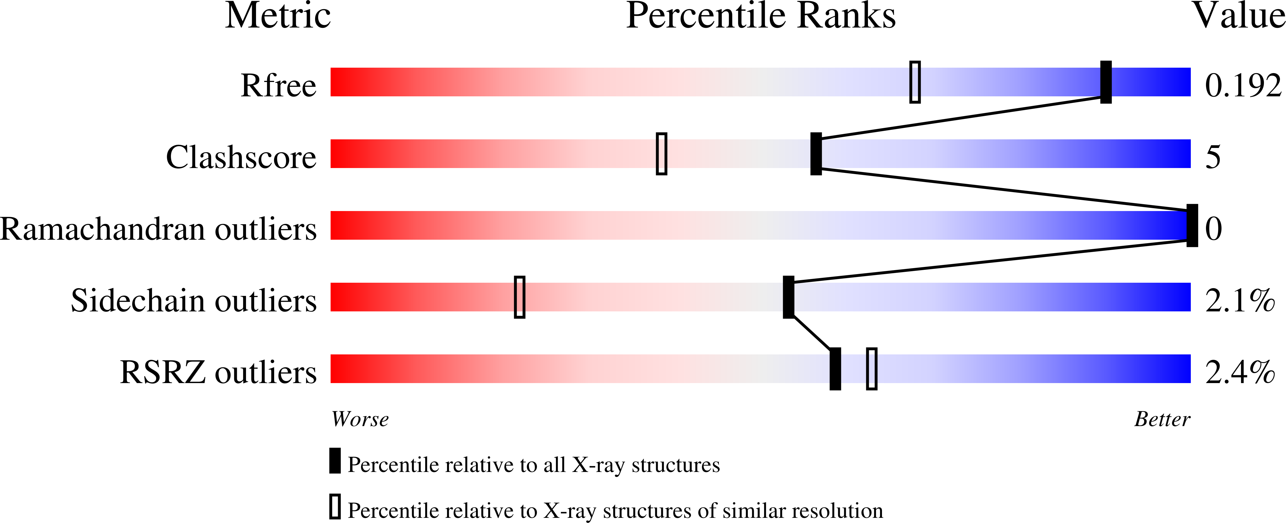
R-Value Free:
0.19
R-Value Work:
0.15
R-Value Observed:
0.15
Space Group:
C 1 2 1