8SAO image
Entry Detail
PDB ID:
8SAO
Title:
Crystal structure of class III lanthipeptide synthetase ThurKC in complex with ThurA1 leader peptide
Biological Source:
PDB Version:
Deposition Date:
2023-04-01
Release Date:
2023-11-01
Method Details:
Experimental Method:
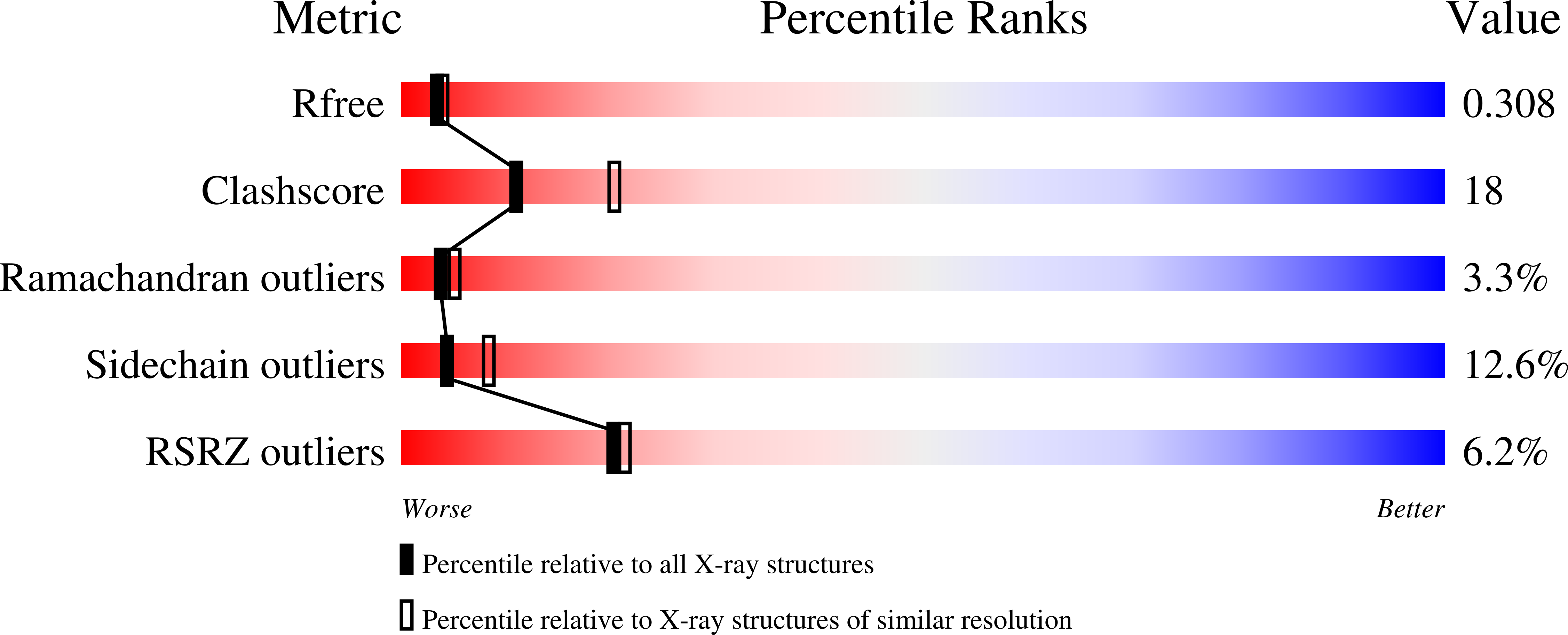
Resolution:
2.50 Å
R-Value Free:
0.30
R-Value Work:
0.24
Space Group:
P 1 21 1
Macromolecular Entities
Polymer Type:polypeptide(L)
Description:Class III lanthipeptide
Chain IDs:A, B (auth: C)
Chain Length:14
Number of Molecules:2
Biological Source:Bacillus thuringiensis serovar andalousiensis
Polymer Type:polypeptide(L)
Description:Class III lanthionine synthetase LanKC
Chain IDs:C (auth: B), D
Chain Length:859
Number of Molecules:2
Biological Source:Bacillus thuringiensis serovar andalousiensis
Ligand Molecules
Primary Citation
Structure and Function of a Class III Metal-Independent Lanthipeptide Synthetase.
Acs Cent.Sci. 9 1944 1956 (2023)
PMID: 37901177 DOI: 10.1021/acscentsci.3c00484

Abstact

In bacteria, Ser/Thr protein kinase-like sequences are found as part of large multidomain polypeptides that biosynthesize lanthipeptides, a class of natural products distinguished by the presence of thioether cross-links. The kinase domain phosphorylates Ser or Thr residues in the peptide substrates. Subsequent β-elimination by a lyase domain yields electrophilic dehydroamino acids, which can undergo cyclase domain-catalyzed cyclization to yield conformationally restricted, bioactive compounds. Here, we reconstitute the biosynthetic pathway for a class III lanthipeptide from Bacillus thuringiensis NRRL B-23139, including characterization of a two-component protease for leader peptide excision. We also describe the first crystal structures of a class III lanthipeptide synthetase, consisting of the lyase, kinase, and cyclase domains, in various states including complexes with its leader peptide and nucleotide. The structure shows interactions between all three domains that result in an active conformation of the kinase domain. Biochemical analysis demonstrates that the three domains undergo movement upon binding of the leader peptide to establish interdomain allosteric interactions that stabilize this active form. These studies inform on the regulatory mechanism of substrate recognition and provide a framework for engineering of variants of biotechnological interest.

Legend

Protein

Chemical

Disease

Primary Citation of related structures