8SA4 image
Entry Detail
PDB ID:
8SA4
EMDB ID:
Keywords:
Title:
Adenosylcobalamin-bound riboswitch dimer, form 3
Biological Source:
PDB Version:
Deposition Date:
2023-03-31
Release Date:
2023-07-26
Method Details:
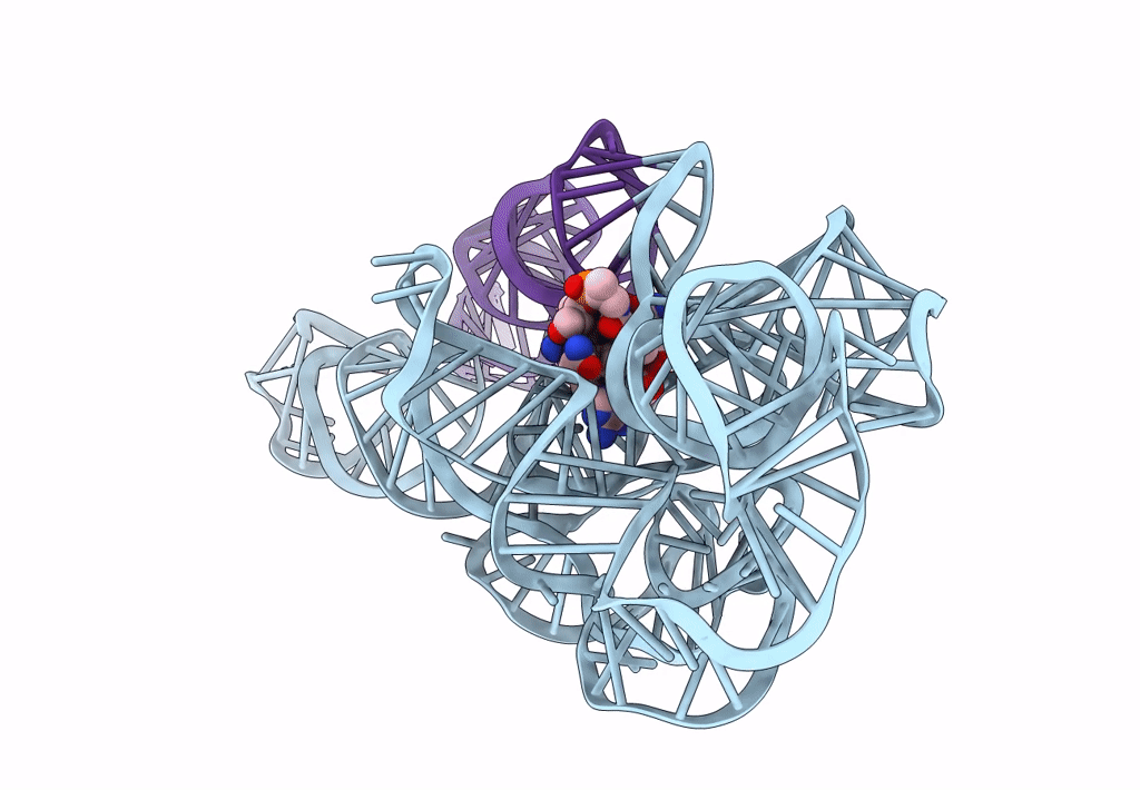
Experimental Method:
Resolution:
3.10 Å
Aggregation State:
PARTICLE
Reconstruction Method:
SINGLE PARTICLE
Macromolecular Entities
Polymer Type:polyribonucleotide
Description:adenosylcobalamin riboswitch form 3
Chain IDs:A, B
Chain Length:210
Number of Molecules:2
Biological Source:Caldanaerobacter subterraneus subsp. tengcongensis
Ligand Molecules
Primary Citation
Capturing heterogeneous conformers of cobalamin riboswitch by cryo-EM.
Nucleic Acids Res. 51 9952 9960 (2023)
PMID: 37534568 DOI: 10.1093/nar/gkad651

Abstact

RNA conformational heterogeneity often hampers its high-resolution structure determination, especially for large and flexible RNAs devoid of stabilizing proteins or ligands. The adenosylcobalamin riboswitch exhibits heterogeneous conformations under 1 mM Mg2+ concentration and ligand binding reduces conformational flexibility. Among all conformers, we determined one apo (5.3 Å) and four holo cryo-electron microscopy structures (overall 3.0-3.5 Å, binding pocket 2.9-3.2 Å). The holo dimers exhibit global motions of helical twisting and bending around the dimer interface. A backbone comparison of the apo and holo states reveals a large structural difference in the P6 extension position. The central strand of the binding pocket, junction 6/3, changes from an 'S'- to a 'U'-shaped conformation to accommodate ligand. Furthermore, the binding pocket can partially form under 1 mM Mg2+ and fully form under 10 mM Mg2+ within the bound-like structure in the absence of ligand. Our results not only demonstrate the stabilizing ligand-induced conformational changes in and around the binding pocket but may also provide further insight into the role of the P6 extension in ligand binding and selectivity.

Legend

Protein

Chemical

Disease

Primary Citation of related structures