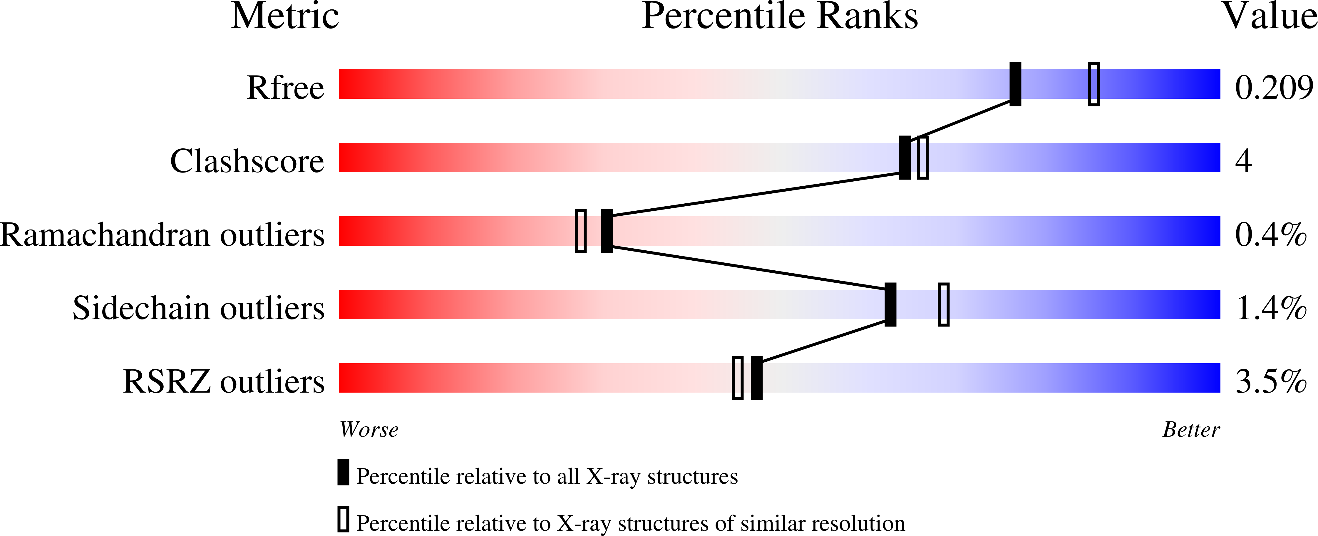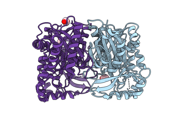
8S81 image
Entry Detail
PDB ID:
8S81
Keywords:
Title:
VirC mutant C114A-Q334A-R335A-R338A
Biological Source:
Source Organism:
PDB Version:
Deposition Date:
2024-03-05
Release Date:
2024-11-13
Method Details:
Experimental Method:
Resolution:
1.99 Å
R-Value Free:
0.20
R-Value Work:
0.17
R-Value Observed:
0.17
Space Group:
H 3 2
Macromolecular Entities
Polymer Type:polypeptide(L)
Description:HMG-CoA synthase-like protein
Chain IDs:A, B
Chain Length:421
Number of Molecules:2
Biological Source:Streptomyces virginiae
Modified Residue
Compound ID Chain ID Parent Comp ID Details 2D Image
MSE A MET modified residue
Ligand Molecules
Primary Citation
Structural Plasticity within 3-Hydroxy-3-Methylglutaryl Synthases Catalyzing the First Step of beta-Branching in Polyketide Biosynthesis Underpins a Dynamic Mechanism of Substrate Accommodation.
Jacs Au 4 3833 3847 (2024)
PMID: 39483223 DOI: 10.1021/jacsau.4c00477

Abstact

Understanding how enzymes have been repurposed by evolution to carry out new functions is a key goal of mechanistic enzymology. In this study we aimed to identify the adaptations required to allow the 3-hydroxy-3-methylglutaryl (HMG)-CoA synthase (HMGCS) enzymes of primary isoprenoid assembly to function in specialized polyketide biosynthetic pathways, where they initiate β-branching. This role notably necessitates that the HMG synthases (HMGSs) act on substrates tethered to noncatalytic acyl carrier protein (ACP) domains instead of coenzyme A, and accommodation of substantially larger chains within the active sites. Here, we show using a combination of X-ray crystallography and small-angle X-ray scattering, that a model HMGS from the virginiamycin system exhibits markedly increased flexibility relative to its characterized HMGCS counterparts. This mobility encompasses multiple secondary structural elements that define the dimensions and chemical nature of the active site, as well the catalytic residues themselves. This result was unexpected given the well-ordered character of the HMGS within the context of an HMGS/ACP complex, but analysis by synchrotron radiation circular dichroism demonstrates that this interaction leads to increased HMGS folding. This flexible to more rigid transition is notably not accounted for by AlphaFold2, which yielded a structural model incompatible with binding of the native substrates. Taken together, these results illustrate the continued necessity of an integrative structural biology approach combining crystallographic and solution-phase data for elucidating the mechanisms underlying enzyme remodeling, information which can inform strategies to replicate such evolution effectively in the laboratory.

Legend

Protein

Chemical

Disease

Primary Citation of related structures