Deposition Date
2024-01-05
Release Date
2024-04-24
Last Version Date
2024-04-24
Entry Detail
PDB ID:
8RM7
Keywords:
Title:
Crystal Structure of Human Androgen Receptor DNA Binding Domain Bound to its Response Element: MMTV-177 GRE/ARE
Biological Source:
Source Organism(s):
Homo sapiens (Taxon ID: 9606)
Expression System(s):
Method Details:
Experimental Method:
Resolution:
2.25 Å
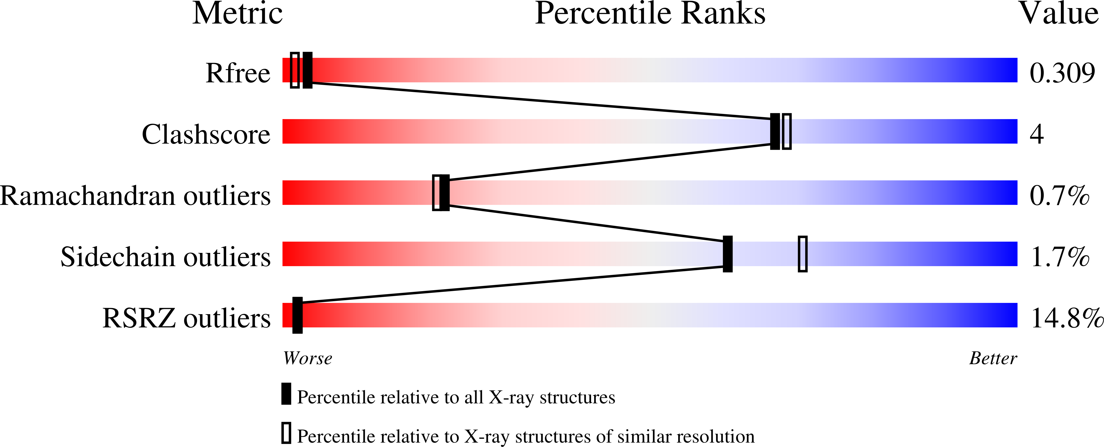
R-Value Free:
0.31
R-Value Work:
0.26
R-Value Observed:
0.26
Space Group:
C 1 2 1