8RFU image
Entry Detail
PDB ID:
8RFU
Keywords:
Title:
Low pH (5.5) nitrite-bound MSOX movie series dataset 40 of the copper nitrite reductase (NirK) from Bradyrhizobium japonicum USDA110 [24.4 MGy] - water ligand + decarboxylated AspCAT (final)
Biological Source:
PDB Version:
Deposition Date:
2023-12-13
Release Date:
2024-07-24
Method Details:
Experimental Method:
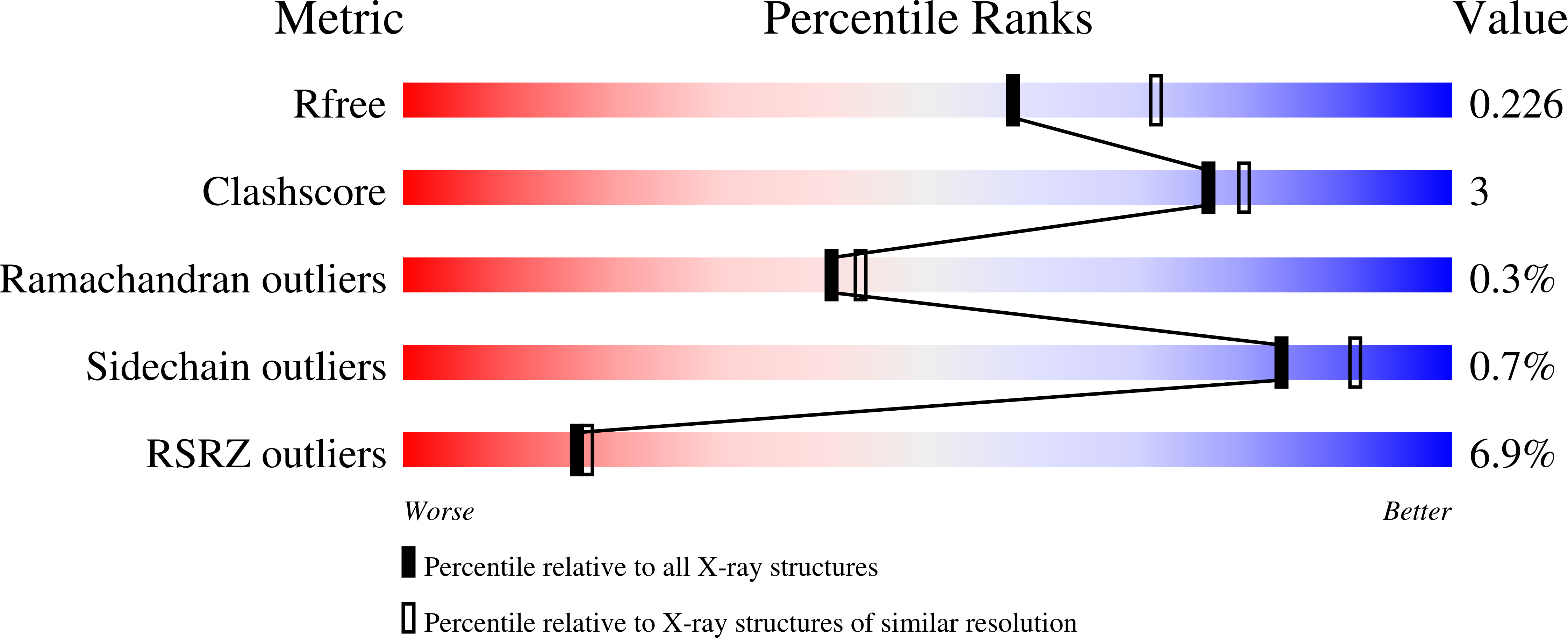
Resolution:
2.18 Å
R-Value Free:
0.22
R-Value Work:
0.18
R-Value Observed:
0.18
Space Group:
P 63
Macromolecular Entities
Polymer Type:polypeptide(L)
Description:Copper-containing nitrite reductase
Chain IDs:A
Chain Length:340
Number of Molecules:1
Biological Source:Bradyrhizobium diazoefficiens USDA 110
Peptide-like Molecules
PRD_900003
Primary Citation
Spectroscopically Validated pH-dependent MSOX Movies Provide Detailed Mechanism of Copper Nitrite Reductases.
J.Mol.Biol. 436 168706 168706 (2024)
PMID: 39002715 DOI: 10.1016/j.jmb.2024.168706

Abstact

Copper nitrite reductases (CuNiRs) exhibit a strong pH dependence of their catalytic activity. Structural movies can be obtained by serially recording multiple structures (frames) from the same spot of a crystal using the MSOX serial crystallography approach. This method has been combined with on-line single crystal optical spectroscopy to capture the pH-dependent structural changes that accompany during turnover of CuNiRs from two Rhizobia species. The structural movies, initiated by the redox activation of a type-1 copper site (T1Cu) via X-ray generated photoelectrons, have been obtained for the substrate-free and substrate-bound states at low (high enzymatic activity) and high (low enzymatic activity) pH. At low pH, formation of the product nitric oxide (NO) is complete at the catalytic type-2 copper site (T2Cu) after a dose of 3 MGy (frame 5) with full bleaching of the T1Cu ligand-to-metal charge transfer (LMCT) 455 nm band (S(σ)Cys → T1Cu2+) which in itself indicates the electronic route of proton-coupled electron transfer (PCET) from T1Cu to T2Cu. In contrast at high pH, the changes in optical spectra are relatively small and the formation of NO is only observed in later frames (frame 15 in Br2DNiR, 10 MGy), consistent with the loss of PCET required for catalysis. This is accompanied by decarboxylation of the catalytic AspCAT residue, with CO2 trapped in the catalytic pocket.

Legend

Protein

Chemical

Disease

Primary Citation of related structures