Deposition Date
2023-12-10
Release Date
2024-01-31
Last Version Date
2024-01-31
Entry Detail
PDB ID:
8RE2
Keywords:
Title:
Crystal Structure determination of Dye-decolorizing Peroxidase (DyP) from Deinoccoccus radiodurans
Biological Source:
Source Organism(s):
Deinococcus radiodurans (Taxon ID: 1299)
Expression System(s):
Method Details:
Experimental Method:
Resolution:
2.20 Å
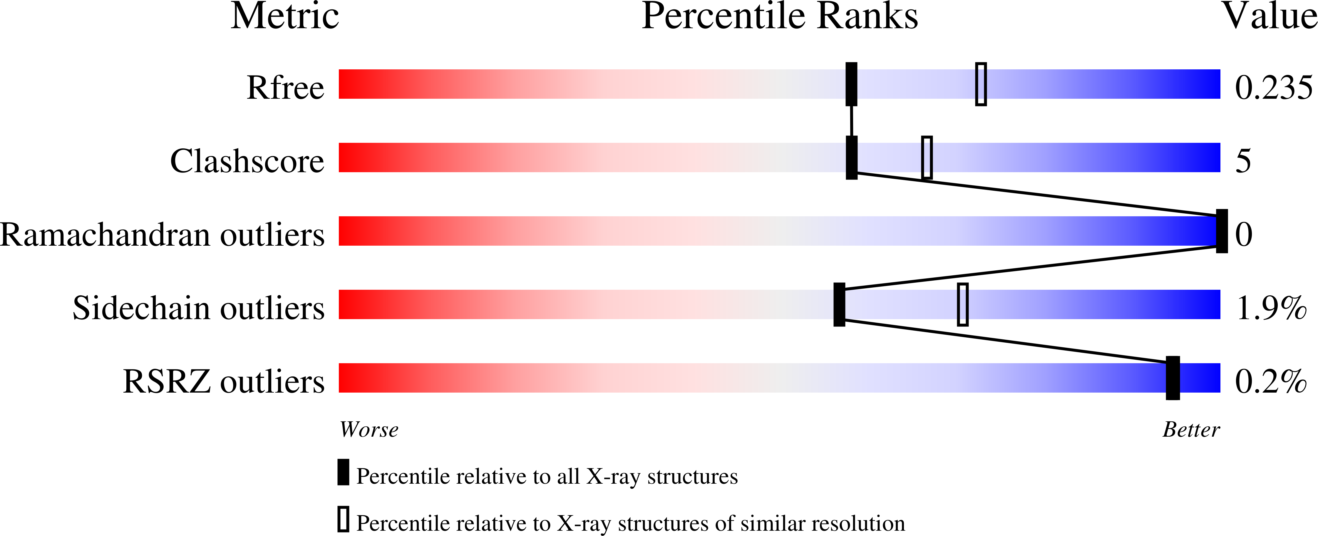
R-Value Free:
0.23
R-Value Work:
0.18
R-Value Observed:
0.18
Space Group:
P 32