
Deposition Date
2023-12-06
Release Date
2024-06-26
Last Version Date
2024-11-20
Entry Detail
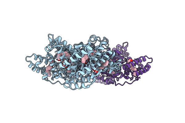
PDB ID:
8RCP
Keywords:
Title:
Structure of Human Serum Albumin in complex with Myristic Acid
Biological Source:
Source Organism(s):
Homo sapiens (Taxon ID: 9606)
Method Details:
Experimental Method:
Resolution:
1.90 Å
R-Value Free:
0.26
R-Value Work:
0.22
R-Value Observed:
0.22
Space Group:
P 1 21 1
