Deposition Date
2023-12-04
Release Date
2024-12-11
Last Version Date
2025-06-04
Entry Detail
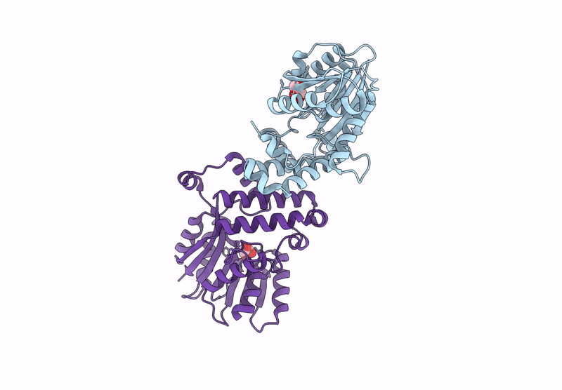
PDB ID:
8RBE
Keywords:
Title:
Crystal structure of Mycobacterium tuberculosis MmaA1 in apo form
Biological Source:
Source Organism(s):
Mycobacterium tuberculosis (Taxon ID: 1773)
Expression System(s):
Method Details:
Experimental Method:
Resolution:
1.90 Å
R-Value Free:
0.20
R-Value Work:
0.17
R-Value Observed:
0.17
Space Group:
P 1 21 1