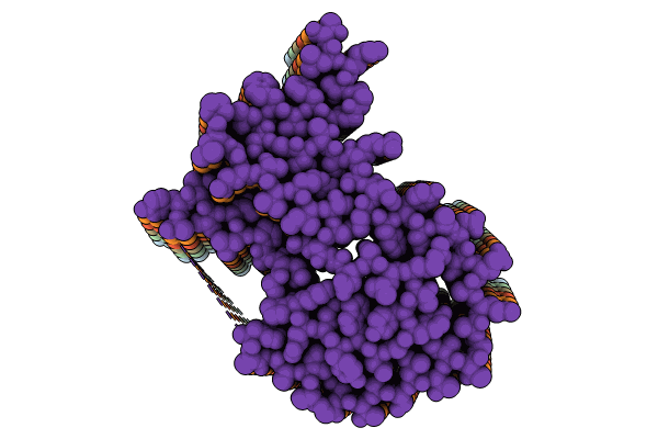
8R47 image
Deposition Date 2023-11-13
Release Date 2024-06-26
Last Version Date 2024-10-23
Entry Detail
PDB ID:
8R47
Keywords:
Title:
AL amyloid fibril from the FOR010 light chain
Biological Source:
Source Organism:
Homo sapiens (Taxon ID: 9606)
Method Details:
Experimental Method:
Resolution:
2.25 Å
Aggregation State:
HELICAL ARRAY
Reconstruction Method:
HELICAL
Macromolecular Entities
Polymer Type:polypeptide(L)
Molecule:lambda 3 immunoglobulin light chain fragment, residues 4-116
Chain IDs:A, B, C, D, E, F
Chain Length:113
Number of Molecules:6
Biological Source:Homo sapiens
Ligand Molecules
Primary Citation
Light chain mutations contribute to defining the fibril morphology in systemic AL amyloidosis.
Nat Commun 15 5121 5121 (2024)
PMID: 38879609 DOI: 10.1038/s41467-024-49520-6

Abstact

Systemic AL amyloidosis is one of the most frequently diagnosed forms of systemic amyloidosis. It arises from mutational changes in immunoglobulin light chains. To explore whether these mutations may affect the structure of the formed fibrils, we determine and compare the fibril structures from several patients with cardiac AL amyloidosis. All patients are affected by light chains that contain an IGLV3-19 gene segment, and the deposited fibrils differ by the mutations within this common germ line background. Using cryo-electron microscopy, we here find different fibril structures in each patient. These data establish that the mutations of amyloidogenic light chains contribute to defining the fibril architecture and hence the structure of the pathogenic agent.

Legend

Protein

Chemical

Disease

Primary Citation of related structures