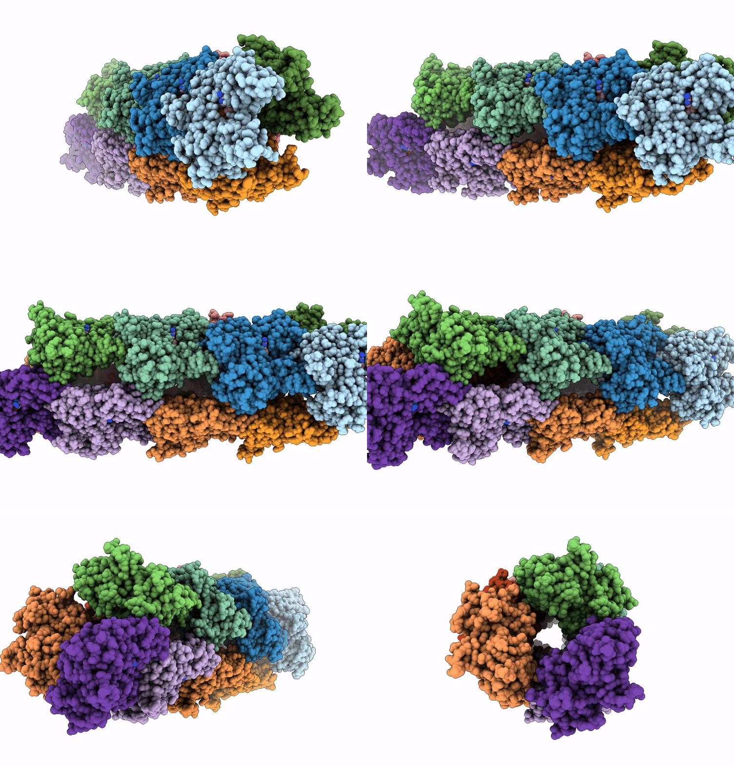
8R2N image
Entry Detail
PDB ID:
8R2N
EMDB ID:
Title:
Structure of the BeeR filament
Biological Source:
Source Organism:
Host Organism:
PDB Version:
Deposition Date:
2023-11-07
Release Date:
2025-03-05
Method Details:
Experimental Method:
Resolution:
3.10 Å
Aggregation State:
FILAMENT
Reconstruction Method:
HELICAL
Macromolecular Entities
Polymer Type:polypeptide(L)
Description:Actin/actin family protein
Chain IDs:A, B, C, D, E, F, G, H, I, J, K, L
Chain Length:386
Number of Molecules:12
Biological Source:Opitutus terrae
Primary Citation
A family of bacterial actin homologs forms a three-stranded tubular structure.
Proc.Natl.Acad.Sci.USA 122 e2500913122 e2500913122 (2025)
PMID: 40073056 DOI: 10.1073/pnas.2500913122

Abstact

The cytoskeleton is crucial for cell organization and movement. In Eukaryotes, it largely consists of the protein actin, that forms a double-stranded linear filamentous structure in the presence of ATP and disassemble upon ATP hydrolysis. Bacteria also possess actin homologs, that drive fundamental cellular processes, including cell division, shape maintenance, and DNA segregation. Like eukaryotic actin, bacterial actins assemble into dynamic polymers upon ATP binding, however variation in interactions between strands gives rise to striking diversity of filament architectures. Here, we report a family of bacterial actins of unknown function, conserved among the Verrucomicrobiota phylum, which assembles into a unique tubular structure in the presence of ATP. A cryo-EM structure of the filaments reveals that it consists of three strands, unlike other described bacterial actin structures. This architecture provides further insights into the organization of actin-like filaments and has implications for understanding the diversity and evolution of the bacterial cytoskeleton.

Legend

Protein

Chemical

Disease

Primary Citation of related structures