8R2C image
Deposition Date 2023-11-03
Release Date 2024-02-28
Last Version Date 2024-05-01
Entry Detail
PDB ID:
8R2C
Title:
Crystal structure of the Vint domain from Tetrahymena thermophila
Biological Source:
Source Organism:
Host Organism:
Method Details:
Experimental Method:
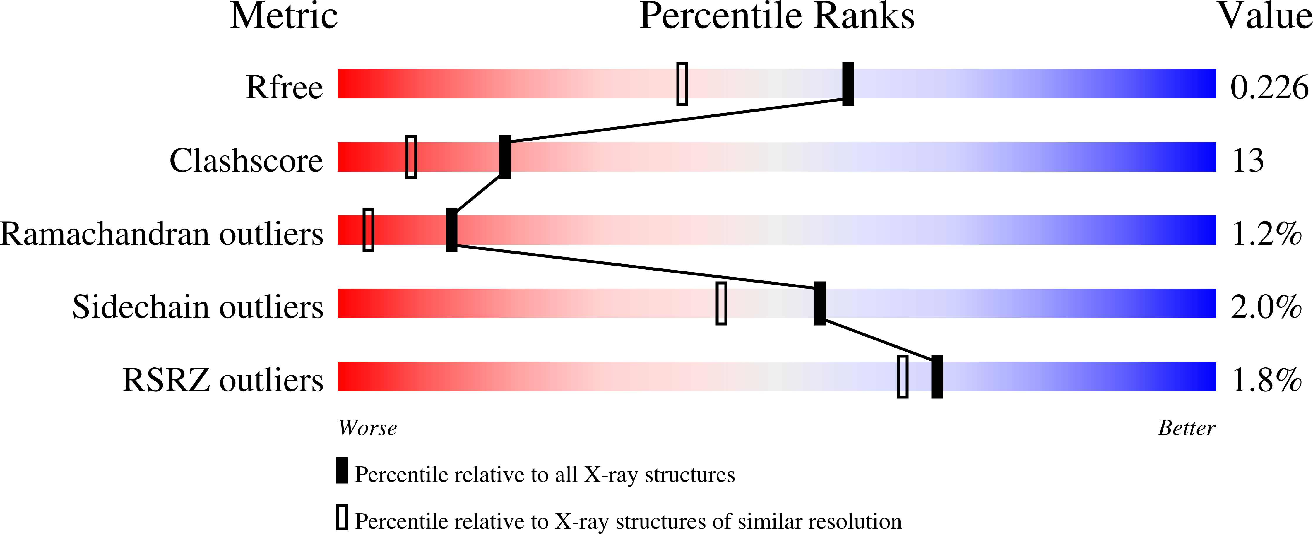
Resolution:
1.80 Å
R-Value Free:
0.22
R-Value Work:
0.18
R-Value Observed:
0.18
Space Group:
P 21 21 21
Macromolecular Entities
Structures with similar UniProt ID
Protein Blast
Polymer Type:polypeptide(L)
Molecule:von willebrand factor type A (VWA) domain was originally protein
Gene (Uniprot):TTHERM_00471620
Chain IDs:A
Chain Length:169
Number of Molecules:1
Biological Source:Tetrahymena thermophila SB210
Primary Citation
The three-dimensional structure of the Vint domain from Tetrahymena thermophila suggests a ligand-regulated cleavage mechanism by the HINT fold.
Febs Lett. 598 864 874 (2024)
PMID: 38351630 DOI: 10.1002/1873-3468.14817

Abstact

Vint proteins have been identified in unicellular metazoans as a novel hedgehog-related gene family, merging the von Willebrand factor type A domain and the Hedgehog/INTein (HINT) domains. We present the first three-dimensional structure of the Vint domain from Tetrahymena thermophila corresponding to the auto-processing domain of hedgehog proteins, shedding light on the unique features, including an adduct recognition region (ARR). Our results suggest a potential binding between the ARR and sulfated glycosaminoglycans like heparin sulfate. Moreover, we uncover a possible regulatory role of the ARR in the auto-processing by Vint domains, expanding our understanding of the HINT domain evolution and their use in biotechnological applications. Vint domains might have played a crucial role in the transition from unicellular to multicellular organisms.

Legend

Protein

Chemical

Disease

Primary Citation of related structures