Deposition Date
2023-10-31
Release Date
2025-05-14
Last Version Date
2025-07-16
Entry Detail
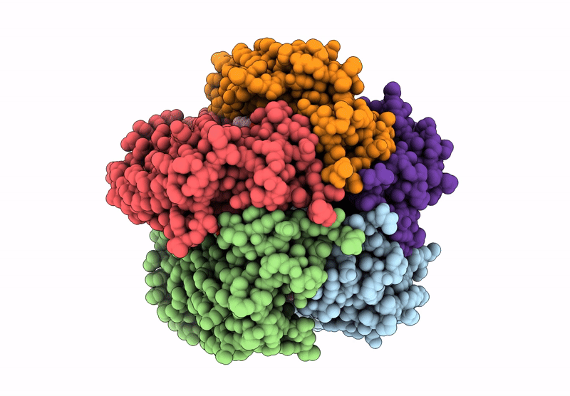
PDB ID:
8R0O
Keywords:
Title:
Cryo-EM structure of the microbial rhodopsin CryoR1 at pH 10.5 in detergent in the M state
Biological Source:
Source Organism(s):
Cryobacterium levicorallinum (Taxon ID: 995038)
Expression System(s):
Method Details:
Experimental Method:
Resolution:
2.30 Å
Aggregation State:
PARTICLE
Reconstruction Method:
SINGLE PARTICLE