8QZZ image
Deposition Date 2023-10-30
Release Date 2024-03-20
Last Version Date 2024-04-10
Entry Detail
PDB ID:
8QZZ
Keywords:
Title:
Crystal structure of human eIF2 alpha-gamma complexed with PPP1R15A_420-452
Biological Source:
Source Organism:
Homo sapiens (Taxon ID: 9606)
Host Organism:
Method Details:
Experimental Method:
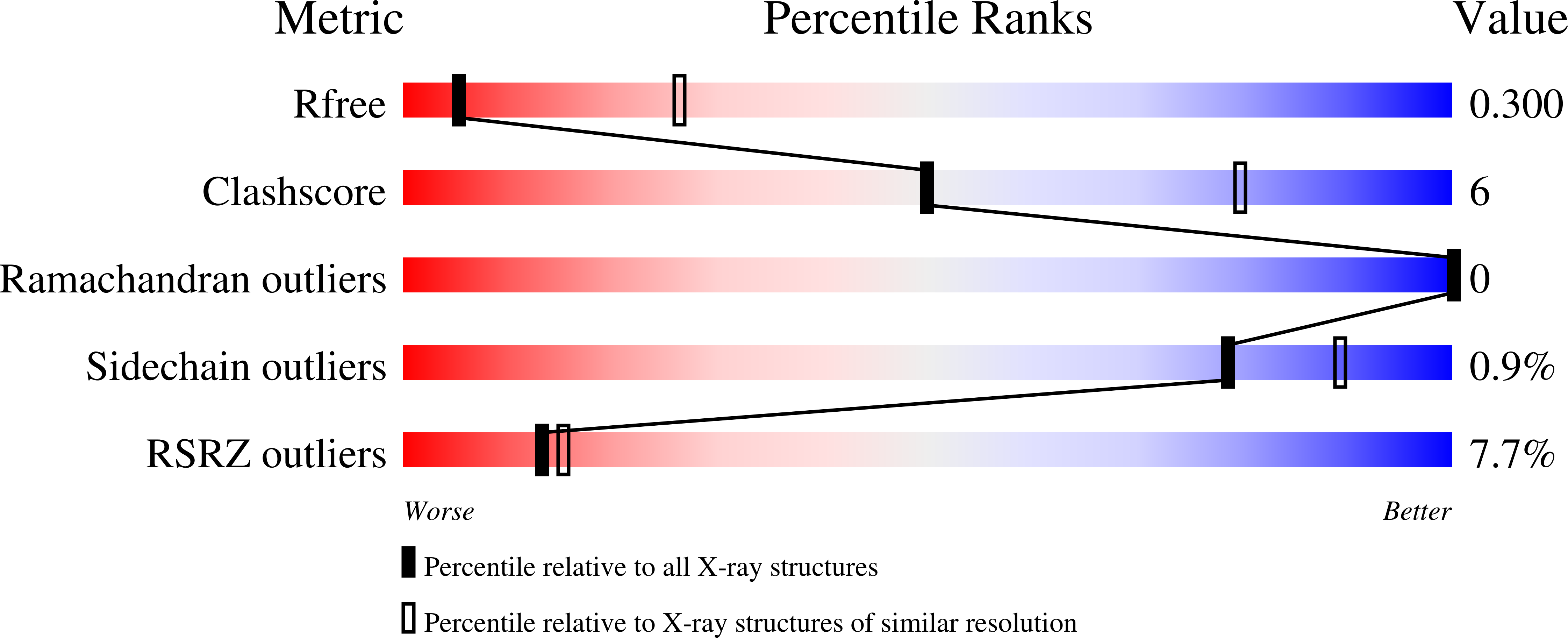
Resolution:
3.35 Å
R-Value Free:
0.29
R-Value Work:
0.24
R-Value Observed:
0.24
Space Group:
P 41 21 2
Macromolecular Entities
Structures with similar UniProt ID
Protein Blast
Polymer Type:polypeptide(L)
Molecule:Eukaryotic translation initiation factor 2 subunit 3
Gene (Uniprot):EIF2S3
Chain IDs:A
Chain Length:472
Number of Molecules:1
Biological Source:Homo sapiens
Structures with similar UniProt ID
Protein Blast
Polymer Type:polypeptide(L)
Molecule:Eukaryotic translation initiation factor 2 subunit 1
Gene (Uniprot):EIF2S1
Chain IDs:B
Chain Length:315
Number of Molecules:1
Biological Source:Homo sapiens
Structures with similar UniProt ID
Protein Blast
Polymer Type:polypeptide(L)
Molecule:Protein phosphatase 1 regulatory subunit 15A
Gene (Uniprot):PPP1R15A
Chain IDs:C
Chain Length:33
Number of Molecules:1
Biological Source:Homo sapiens
Ligand Molecules
Primary Citation
Substrate recruitment via eIF2 gamma enhances catalytic efficiency of a holophosphatase that terminates the integrated stress response.
Proc.Natl.Acad.Sci.USA 121 e2320013121 e2320013121 (2024)
PMID: 38547060 DOI: 10.1073/pnas.2320013121

Abstact

Dephosphorylation of pSer51 of the α subunit of translation initiation factor 2 (eIF2αP) terminates signaling in the integrated stress response (ISR). A trimeric mammalian holophosphatase comprised of a protein phosphatase 1 (PP1) catalytic subunit, the conserved C-terminally located ~70 amino acid core of a substrate-specific regulatory subunit (PPP1R15A/GADD34 or PPP1R15B/CReP) and G-actin (an essential cofactor) efficiently dephosphorylate eIF2αP in vitro. Unlike their viral or invertebrate counterparts, with whom they share the conserved 70 residue core, the mammalian PPP1R15s are large proteins of more than 600 residues. Genetic and cellular observations point to a functional role for regions outside the conserved core of mammalian PPP1R15A in dephosphorylating its natural substrate, the eIF2 trimer. We have combined deep learning technology, all-atom molecular dynamics simulations, X-ray crystallography, and biochemistry to uncover binding of the γ subunit of eIF2 to a short helical peptide repeated four times in the functionally important N terminus of human PPP1R15A that extends past its conserved core. Binding entails insertion of Phe and Trp residues that project from one face of an α-helix formed by the conserved repeats of PPP1R15A into a hydrophobic groove exposed on the surface of eIF2γ in the eIF2 trimer. Replacing these conserved Phe and Trp residues with Ala compromises PPP1R15A function in cells and in vitro. These findings suggest mechanisms by which contacts between a distant subunit of eIF2 and elements of PPP1R15A distant to the holophosphatase active site contribute to dephosphorylation of eIF2αP by the core PPP1R15 holophosphatase and to efficient termination of the ISR in mammals.

Legend

Protein

Chemical

Disease

Primary Citation of related structures
Feedback Form
Name
Email
Institute
Feedback