8QV3 image
Deposition Date 2023-10-17
Release Date 2024-04-24
Last Version Date 2024-07-31
Entry Detail
PDB ID:
8QV3
Keywords:
Title:
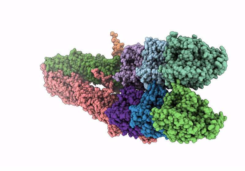
Structure of the y-Tubulin Small Complex (yTuSC) as part of the native y-Tubulin Ring Complex (yTuRC) capping microtubule minus ends at the spindle pole body
Biological Source:
Source Organism:
Method Details:
Experimental Method:
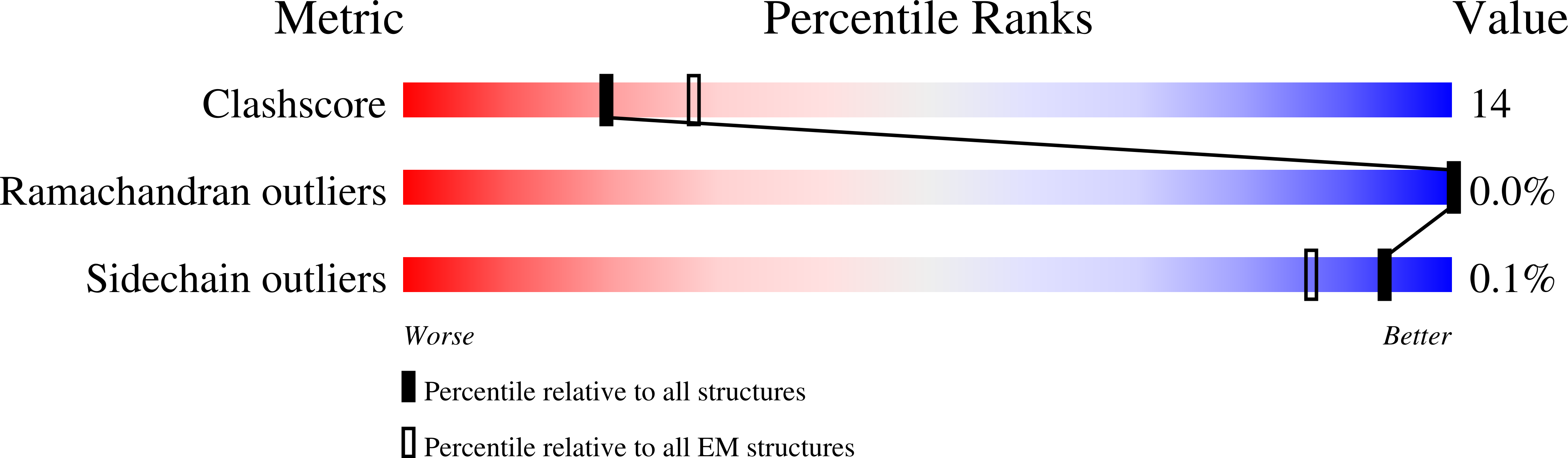
Resolution:
8.20 Å
Aggregation State:
PARTICLE
Reconstruction Method:
SUBTOMOGRAM AVERAGING
Macromolecular Entities
Structures with similar UniProt ID
Protein Blast
Polymer Type:polypeptide(L)
Molecule:Tubulin alpha-1 chain
Gene (Uniprot):TUB1
Chain IDs:G (auth: Ad), H (auth: Ac)
Chain Length:447
Number of Molecules:2
Biological Source:Saccharomyces cerevisiae
Structures with similar UniProt ID
Protein Blast
Polymer Type:polypeptide(L)
Molecule:Tubulin beta chain
Chain IDs:I (auth: Bd), J (auth: Bc)
Chain Length:457
Number of Molecules:2
Biological Source:Saccharomyces cerevisiae
Structures with similar UniProt ID
Protein Blast
Polymer Type:polypeptide(L)
Molecule:Spindle pole body component
Chain IDs:C (auth: E)
Chain Length:823
Number of Molecules:1
Biological Source:Saccharomyces cerevisiae
Structures with similar UniProt ID
Protein Blast
Polymer Type:polypeptide(L)
Molecule:Spindle pole body component
Chain IDs:D (auth: F)
Chain Length:846
Number of Molecules:1
Biological Source:Saccharomyces cerevisiae
Structures with similar UniProt ID
Protein Blast
Polymer Type:polypeptide(L)
Molecule:Spindle pole body component 110
Chain IDs:E (auth: Sc), F (auth: Sd)
Chain Length:944
Number of Molecules:2
Biological Source:Saccharomyces cerevisiae
Polymer Type:polypeptide(L)
Molecule:Unknown protein
Chain IDs:K (auth: Uc)
Chain Length:67
Number of Molecules:1
Biological Source:Saccharomyces cerevisiae
Polymer Type:polypeptide(L)
Molecule:Unknown protein
Chain IDs:L (auth: Ud)
Chain Length:62
Number of Molecules:1
Biological Source:Saccharomyces cerevisiae
Structures with similar UniProt ID
Protein Blast
Polymer Type:polypeptide(L)
Molecule:Tubulin gamma chain
Chain IDs:A (auth: c), B (auth: d)
Chain Length:473
Number of Molecules:2
Biological Source:Saccharomyces cerevisiae
Ligand Molecules
Primary Citation
Structure of the native gamma-tubulin ring complex capping spindle microtubules.
Nat.Struct.Mol.Biol. 31 1134 1144 (2024)
PMID: 38609662 DOI: 10.1038/s41594-024-01281-y

Abstact

Microtubule (MT) filaments, composed of α/β-tubulin dimers, are fundamental to cellular architecture, function and organismal development. They are nucleated from MT organizing centers by the evolutionarily conserved γ-tubulin ring complex (γTuRC). However, the molecular mechanism of nucleation remains elusive. Here we used cryo-electron tomography to determine the structure of the native γTuRC capping the minus end of a MT in the context of enriched budding yeast spindles. In our structure, γTuRC presents a ring of γ-tubulin subunits to seed nucleation of exclusively 13-protofilament MTs, adopting an active closed conformation to function as a perfect geometric template for MT nucleation. Our cryo-electron tomography reconstruction revealed that a coiled-coil protein staples the first row of α/β-tubulin of the MT to alternating positions along the γ-tubulin ring of γTuRC. This positioning of α/β-tubulin onto γTuRC suggests a role for the coiled-coil protein in augmenting γTuRC-mediated MT nucleation. Based on our results, we describe a molecular model for budding yeast γTuRC activation and MT nucleation.

Legend

Protein

Chemical

Disease

Primary Citation of related structures