8QQK image
Deposition Date 2023-10-05
Release Date 2024-04-24
Last Version Date 2024-05-01
Entry Detail
PDB ID:
8QQK
Title:
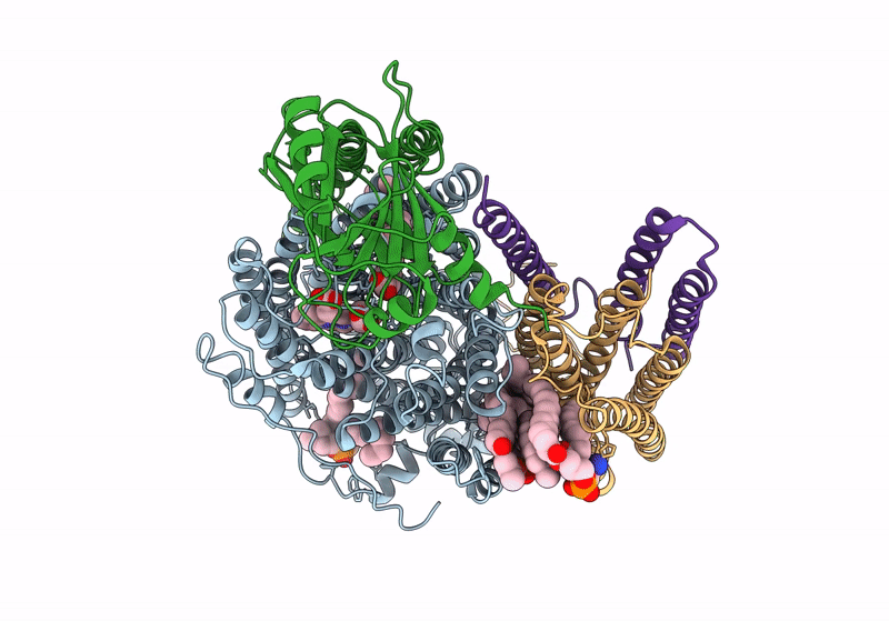
Cryo-EM structure of E. coli cytochrome bo3 quinol oxidase assembled in peptidiscs
Biological Source:
Source Organism:
Method Details:
Experimental Method:
Resolution:
2.80 Å
Aggregation State:
PARTICLE
Reconstruction Method:
SINGLE PARTICLE
Macromolecular Entities
Polymer Type:polypeptide(L)
Molecule:Cytochrome bo(3) ubiquinol oxidase subunit 1
Gene (Uniprot):cyoB
Chain IDs:A
Chain Length:663
Number of Molecules:1
Biological Source:Escherichia coli BL21(DE3)
Polymer Type:polypeptide(L)
Molecule:Cytochrome bo(3) ubiquinol oxidase subunit 2
Gene (Uniprot):cyoA
Chain IDs:B
Chain Length:315
Number of Molecules:1
Biological Source:Escherichia coli BL21(DE3)
Polymer Type:polypeptide(L)
Molecule:Cytochrome bo(3) ubiquinol oxidase subunit 3
Gene (Uniprot):cyoC
Chain IDs:C
Chain Length:204
Number of Molecules:1
Biological Source:Escherichia coli BL21(DE3)
Polymer Type:polypeptide(L)
Molecule:Cytochrome bo(3) ubiquinol oxidase subunit 4
Gene (Uniprot):cyoD
Chain IDs:D
Chain Length:109
Number of Molecules:1
Biological Source:Escherichia coli BL21(DE3)
Primary Citation
Cryo-EM structure of cytochrome bo 3 quinol oxidase assembled in peptidiscs reveals an "open" conformation for potential ubiquinone-8 release.
Biochim Biophys Acta Bioenerg 1865 149045 149045 (2024)
PMID: 38614453 DOI: 10.1016/j.bbabio.2024.149045

Abstact

Cytochrome bo3 quinol oxidase belongs to the heme‑copper-oxidoreductase (HCO) superfamily, which is part of the respiratory chain and essential for cell survival. While the reaction mechanism of cyt bo3 has been studied extensively over the last decades, specific details about its substrate binding and product release have remained unelucidated due to the lack of structural information. Here, we report a 2.8 Å cryo-electron microscopy structure of cyt bo3 from Escherichia coli assembled in peptidiscs. Our structural model shows a conformation for amino acids 1-41 of subunit I different from all previously published structures while the remaining parts of this enzyme are similar. Our new conformation shows a "U-shape" assembly in contrast to the transmembrane helix, named "TM0", in other reported structural models. However, TM0 blocks ubiquinone-8 (reaction product) release, suggesting that other cyt bo3 conformations should exist. Our structural model presents experimental evidence for an "open" conformation to facilitate substrate/product exchange. This work helps further understand the reaction cycle of this oxidase, which could be a benefit for potential drug/antibiotic design for health science.

Legend

Protein

Chemical

Disease

Primary Citation of related structures