Deposition Date
2023-09-26
Release Date
2023-12-20
Last Version Date
2024-10-16
Entry Detail
PDB ID:
8QND
Keywords:
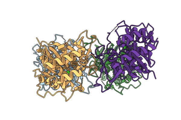
Title:
Crystal structure of the ribonucleoside hydrolase C from Lactobacillus reuteri
Biological Source:
Source Organism(s):
Limosilactobacillus reuteri (Taxon ID: 1598)
Expression System(s):
Method Details:
Experimental Method:
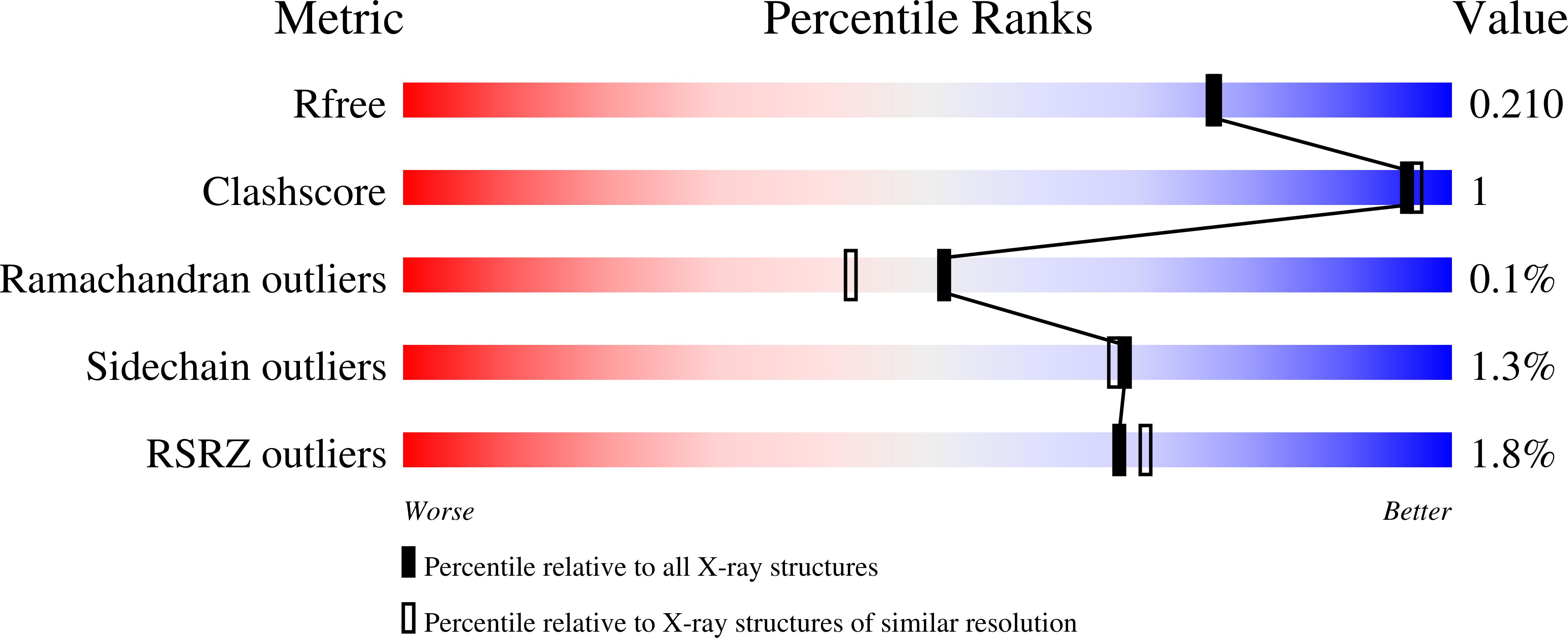
Resolution:
1.90 Å
R-Value Free:
0.20
R-Value Work:
0.16
R-Value Observed:
0.16
Space Group:
P 1 21 1