8QBW image
Deposition Date 2023-08-25
Release Date 2024-09-11
Last Version Date 2025-03-26
Entry Detail
PDB ID:
8QBW
Title:
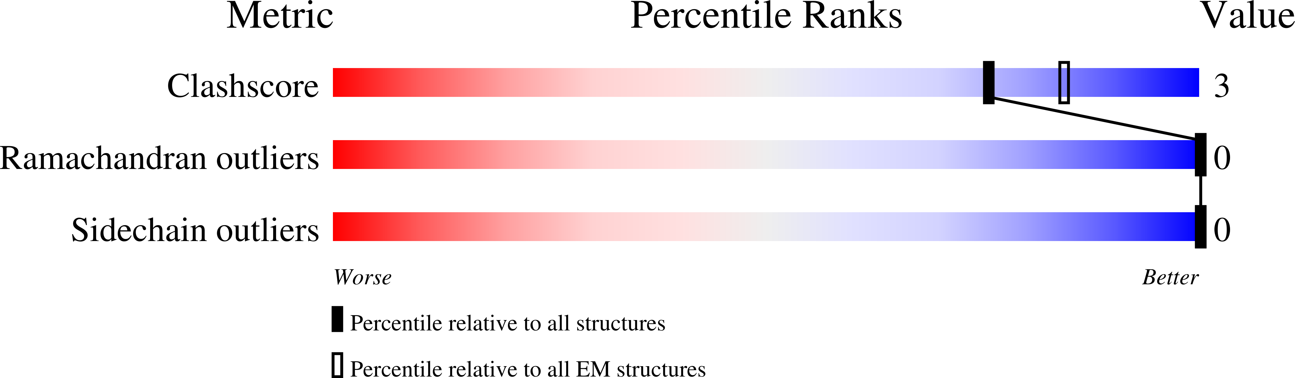
Cryo-EM structure of Vipp1-deltaH6_aa1-219 helical filament with lattice 3 (Vipp1-deltaH6_L3)
Biological Source:
Source Organism:
Host Organism:
Method Details:
Experimental Method:
Resolution:
3.67 Å
Aggregation State:
FILAMENT
Reconstruction Method:
HELICAL
Macromolecular Entities
Polymer Type:polypeptide(L)
Molecule:Phage shock protein A, PspA
Gene (Uniprot):vipp1
Chain IDs:A
Chain Length:219
Number of Molecules:1
Biological Source:Nostoc punctiforme
Ligand Molecules
Primary Citation
Mechanism for Vipp1 spiral formation, ring biogenesis, and membrane repair.
Nat.Struct.Mol.Biol. 32 571 584 (2025)
PMID: 39528797 DOI: 10.1038/s41594-024-01401-8

Abstact

The ESCRT-III-like protein Vipp1 couples filament polymerization with membrane remodeling. It assembles planar sheets as well as 3D rings and helical polymers, all implicated in mitigating plastid-associated membrane stress. The architecture of Vipp1 planar sheets and helical polymers remains unknown, as do the geometric changes required to transition between polymeric forms. Here we show how cyanobacterial Vipp1 assembles into morphologically-related sheets and spirals on membranes in vitro. The spirals converge to form a central ring similar to those described in membrane budding. Cryo-EM structures of helical filaments reveal a close geometric relationship between Vipp1 helical and planar lattices. Moreover, the helical structures reveal how filaments twist-a process required for Vipp1, and likely other ESCRT-III filaments, to transition between planar and 3D architectures. Overall, our results provide a molecular model for Vipp1 ring biogenesis and a mechanism for Vipp1 membrane stabilization and repair, with implications for other ESCRT-III systems.

Legend

Protein

Chemical

Disease

Primary Citation of related structures