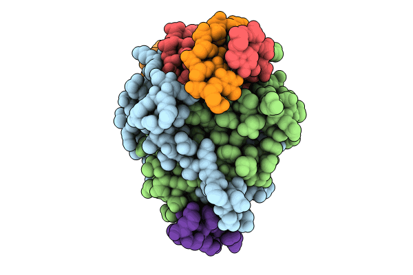
8Q9N image
Entry Detail
PDB ID:
8Q9N
Keywords:
Title:
Crystal Structure of the MADS-box/MEF2 Domain of MEF2D bound to dsDNA and MITR deacetylase binding motif mutant L151V.
Biological Source:
Source Organism:
PDB Version:
Deposition Date:
2023-08-20
Release Date:
2024-06-26
Method Details:
Experimental Method:
Resolution:
1.51 Å
R-Value Free:
0.24
R-Value Work:
0.19
R-Value Observed:
0.19
Space Group:
P 21 21 21
Macromolecular Entities
Polymer Type:polypeptide(L)
Description:MEF2D protein
Chain IDs:A, B
Chain Length:95
Number of Molecules:2
Biological Source:Homo sapiens
Polymer Type:polydeoxyribonucleotide
Description:DNA MADS box
Chain IDs:C (auth: K)
Chain Length:14
Number of Molecules:1
Biological Source:Homo sapiens
Polymer Type:polydeoxyribonucleotide
Description:DNA MADS box
Chain IDs:D (auth: L)
Chain Length:14
Number of Molecules:1
Biological Source:Homo sapiens
Polymer Type:polypeptide(L)
Description:myocyte enhancer-binding factor 2-interacting transcriptional repressor (MITR or HDAC9) L151V mutant peptide: GLU-VAL-LYS-GLN-LYS-LEU-GLN-GLU-PHE-VAL-LEU-SER-LYS
Chain IDs:E (auth: X)
Chain Length:19
Number of Molecules:1
Biological Source:Homo sapiens
Ligand Molecules
Primary Citation
Folding of Class IIa HDAC Derived Peptides into alpha-helices Upon Binding to Myocyte Enhancer Factor-2 in Complex with DNA.
J.Mol.Biol. 436 168541 168541 (2024)
PMID: 38492719 DOI: 10.1016/j.jmb.2024.168541

Abstact

Interaction of transcription factor myocyte enhancer factor-2 (MEF2) family members with class IIa histone deacetylases (HDACs) has been implicated in a wide variety of diseases. Though considerable knowledge on this topic has been accumulated over the years, a high resolution and detailed analysis of the binding mode of multiple class IIa HDAC derived peptides with MEF2D is still lacking. To fulfil this gap, we report here the crystal structure of MEF2D in complex with double strand DNA and four different class IIa HDAC derived peptides, namely HDAC4, HDAC5, HDAC7 and HDAC9. All class IIa HDAC derived peptides form extended amphipathic α-helix structures that fit snugly in the hydrophobic groove of MEF2D domain. Binding mode of class IIa HDAC derived peptides to MEF2D is very similar and occur primarily through nonpolar interactions mediated by highly conserved branched hydrophobic amino acids. Further studies revealed that class IIa HDAC derived peptides are unstructured in solution and appear to adopt a folded α-helix structure only upon binding to MEF2D. Comparison of our peptide-protein complexes with previously characterized structures of MEF2 bound to different co-activators and co-repressors, highlighted both differences and similarities, and revealed the adaptability of MEF2 in protein-protein interactions. The elucidation of the three-dimensional structure of MEF2D in complex with multiple class IIa HDAC derived peptides provide not only a better understanding of the molecular basis of their interactions but also have implications for the development of novel antagonist.

Legend

Protein

Chemical

Disease

Primary Citation of related structures